,.
Invest Ophthalmol Vis Sci. 2020 Jun 3;61(6):33. doi: 10.1167/iovs.61.6.33.
We examined inferior oblique muscles from subjects with over-elevation in adduction for characteristics that might shed light on the potential mechanisms for their abnormal eye position.
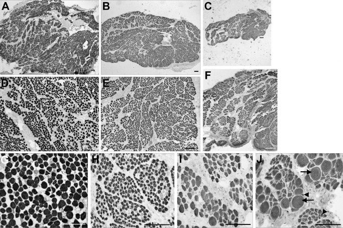
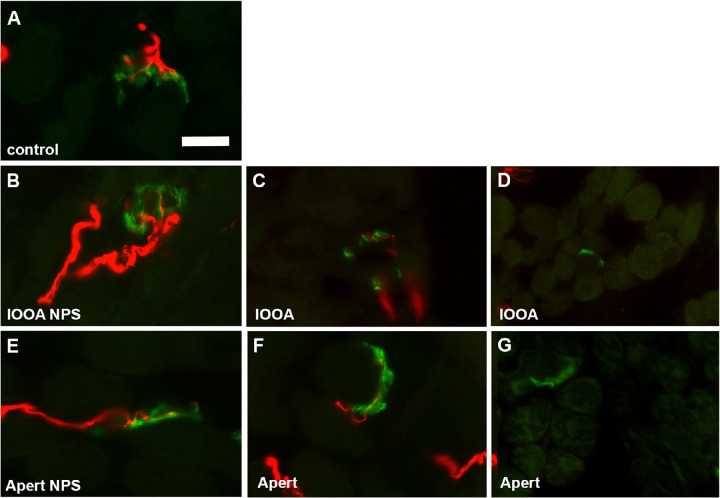
The inferior oblique muscles were obtained at the time of surgery in subjects diagnosed with either primary inferior oblique overaction or Apert syndrome. The muscles were frozen and processed for morphometric analysis of myofiber size, central nucleation, myosin heavy chain (MyHC) isoform expression, nerve density, and numbers of neuromuscular junctions per muscle section.
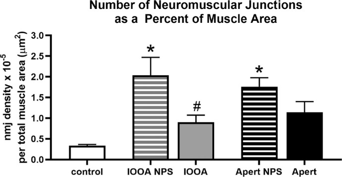
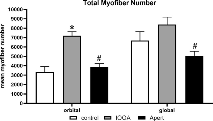
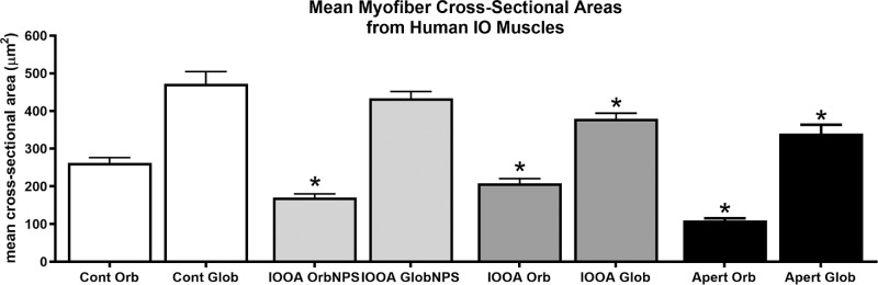
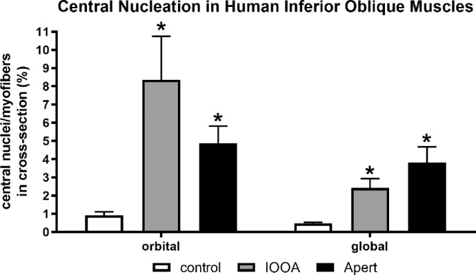
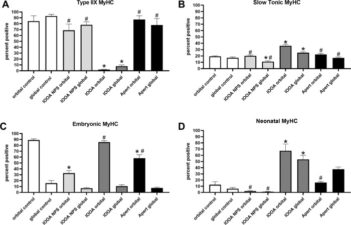
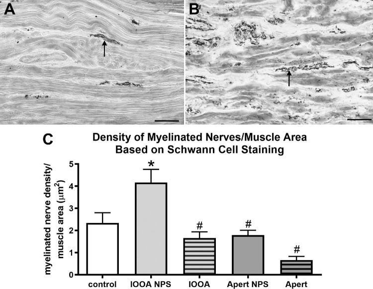
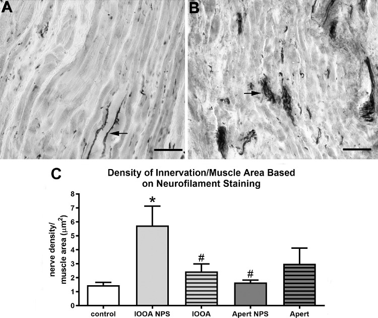
The inferior oblique muscles from subjects with Apert Syndrome were smaller, and had a much more heterogeneous profile relative to myofiber cross-sectional area compared to controls. Increased central nucleation in the Apert syndrome muscles suggested on-going myofiber regeneration or reinnervation over time. Complex changes were seen in the MyHC isoform patterns that would predict slower and more sustained contractions than in the control muscles. Nerve fiber densities were significantly increased compared to controls for the muscles with primary inferior oblique overaction and Apert syndrome that had no prior surgery. The muscles from Apert syndrome subjects as well as those with primary inferior oblique overaction with no prior surgery had significantly elevated numbers of neuromuscular junctions relative to the whole muscle area.
The muscles from both sets of subjects were significantly different from control muscles in a number of properties examined. These data support the view that despite similar manifestations of eye misalignment, the potential mechanism behind the strabismus in these subjects is significantly different.
我们研究了在内收时上抬的患者的下斜肌,以寻找可能揭示其异常眼位潜在机制的特征。
在诊断为原发性下斜肌亢进或 Apert 综合征的患者手术时获取下斜肌。将肌肉冷冻并进行形态计量分析,以测量肌纤维大小、中央核化、肌球蛋白重链(MyHC)同工型表达、神经密度以及每块肌肉切片的运动终板数量。
与对照组相比,Apert 综合征患者的下斜肌较小,且相对于肌纤维横截面积,具有更大的异质性。Apert 综合征肌肉中的中央核化增加表明随着时间的推移,肌纤维不断再生或重新支配。MyHC 同工型模式发生了复杂的变化,预计会产生比对照组更慢和更持久的收缩。与对照组相比,原发性下斜肌亢进和未行手术的 Apert 综合征患者的神经纤维密度显著增加。与整个肌肉面积相比,Apert 综合征患者和未行手术的原发性下斜肌亢进患者的运动终板数量明显升高。
两组患者的肌肉在许多检查的特性上与对照组肌肉明显不同。这些数据支持这样一种观点,即尽管斜视表现相似,但这些患者斜视的潜在机制却大不相同。