Department of Physics, University of York, Heslington YO10 5DD, UK.
Department of Biology, University of York, Heslington YO10 5DD, UK.
Methods. 2021 Sep;193:80-95. doi: 10.1016/j.ymeth.2020.06.007. Epub 2020 Jun 13.
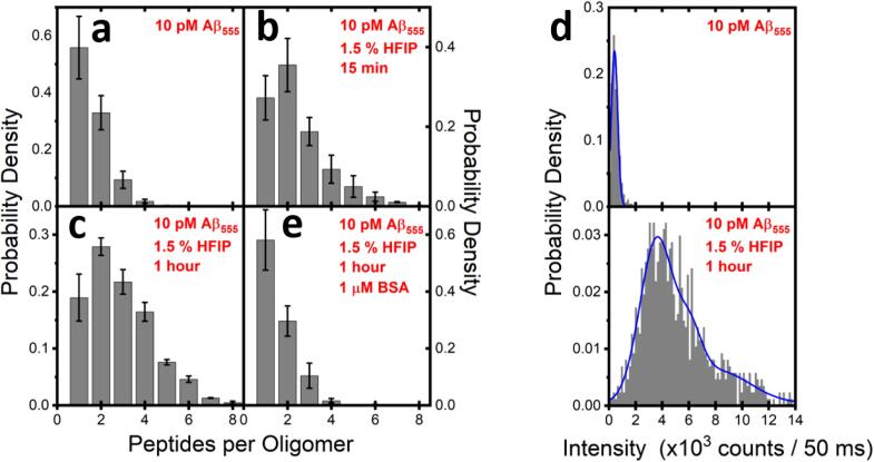
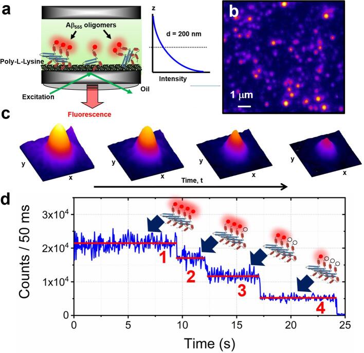
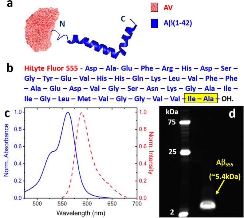
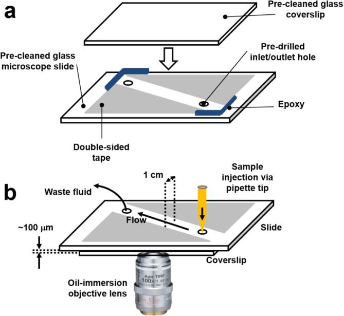
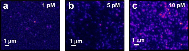
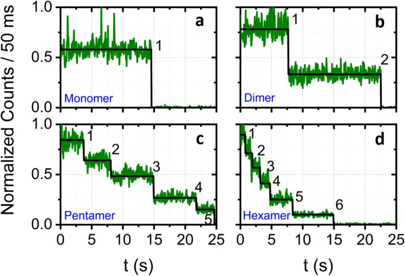
A major hallmark of Alzheimer's disease is the misfolding and aggregation of the amyloid- β peptide (Aβ). While early research pointed towards large fibrillar- and plaque-like aggregates as being the most toxic species, recent evidence now implicates small soluble Aβ oligomers as being orders of magnitude more harmful. Techniques capable of characterizing oligomer stoichiometry and assembly are thus critical for a deeper understanding of the earliest stages of neurodegeneration and for rationally testing next-generation oligomer inhibitors. While the fluorescence response of extrinsic fluorescent probes such as Thioflavin-T have become workhorse tools for characterizing large Aβ aggregates in solution, it is widely accepted that these methods suffer from many important drawbacks, including an insensitivity to oligomeric species. Here, we integrate several biophysics techniques to gain new insight into oligomer formation at the single-molecule level. We showcase single-molecule stepwise photobleaching of fluorescent dye molecules as a powerful method to bypass many of the traditional limitations, and provide a step-by-step guide to implementing the technique in vitro. By collecting fluorescence emission from single Aβ(1-42) peptides labelled at the N-terminal position with HiLyte Fluor 555 via wide-field total internal reflection fluorescence (TIRF) imaging, we demonstrate how to characterize the number of peptides per single immobile oligomer and reveal heterogeneity within sample populations. Importantly, fluorescence emerging from Aβ oligomers cannot be easily investigated using diffraction-limited optical microscopy tools. To assay oligomer activity, we also demonstrate the implementation of another biophysical method involving the ratiometric imaging of Fura-2-AM loaded cells which quantifies the rate of oligomer-induced dysregulation of intracellular Ca homeostasis. We anticipate that the integrated single-molecule biophysics approaches highlighted here will develop further and in principle may be extended to the investigation of other protein aggregation systems under controlled experimental conditions.
阿尔茨海默病的一个主要标志是淀粉样β肽(Aβ)的错误折叠和聚集。虽然早期研究指出大纤维状和斑块状聚集体是最具毒性的物种,但最近的证据表明,小可溶性 Aβ寡聚物的危害要大几个数量级。因此,能够描述寡聚物化学计量和组装的技术对于更深入地了解神经退行性变的早期阶段以及合理测试下一代寡聚物抑制剂至关重要。虽然外源性荧光探针(如硫黄素 T)的荧光响应已成为表征溶液中大 Aβ 聚集体的常用工具,但人们普遍认为这些方法存在许多重要的缺点,包括对寡聚物物种的不敏感。在这里,我们整合了几种生物物理技术,从单分子水平上深入了解寡聚物的形成。我们展示了荧光染料分子的单分子逐步光漂白作为一种强大的方法来绕过许多传统限制,并提供了在体外实施该技术的分步指南。通过使用宽场全内反射荧光(TIRF)成像对 N 端用 HiLyte Fluor 555 标记的单个 Aβ(1-42)肽进行单分子逐步光漂白,我们展示了如何表征每个单固定寡聚物的肽数,并揭示了样品群体内的异质性。重要的是,使用衍射受限的光学显微镜工具很难研究 Aβ 寡聚物的荧光。为了测定寡聚物的活性,我们还展示了另一种生物物理方法的实施情况,该方法涉及负载 Fura-2-AM 的细胞的比率成像,该方法定量测定寡聚物诱导的细胞内 Ca 稳态失调的速率。我们预计,这里强调的集成单分子生物物理方法将进一步发展,并且原则上可以扩展到在受控实验条件下研究其他蛋白质聚集系统。