Division of Nephrology and Hypertension, Department of Medicine, University of Louisville, Louisville, Kentucky
Division of Nephrology and Hypertension, Department of Medicine, University of Louisville, Louisville, Kentucky.
J Am Soc Nephrol. 2020 Aug;31(8):1883-1904. doi: 10.1681/ASN.2019070696. Epub 2020 Jun 19.
The mechanisms leading to extracellular matrix (ECM) replacement of areas of glomerular capillaries in histologic variants of FSGS are unknown. This study used proteomics to test the hypothesis that glomerular ECM composition in collapsing FSGS (cFSGS) differs from that of other variants.
ECM proteins in glomeruli from biopsy specimens of patients with FSGS not otherwise specified (FSGS-NOS) or cFSGS and from normal controls were distinguished and quantified using mass spectrometry, verified and localized using immunohistochemistry (IHC) and confocal microscopy, and assessed for gene expression. The analysis also quantified urinary excretion of ECM proteins and peptides.
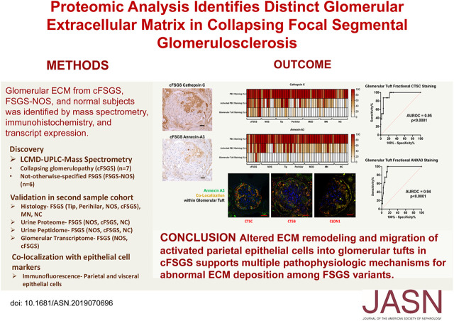
Of 58 ECM proteins that differed in abundance between cFSGS and FSGS-NOS, 41 were more abundant in cFSGS and 17 in FSGS-NOS. IHC showed that glomerular tuft staining for cathepsin B, cathepsin C, and annexin A3 in cFSGS was significantly greater than in other FSGS variants, in minimal change disease, or in membranous nephropathy. Annexin A3 colocalized with cathepsin B and C, claudin-1, phosphorylated ERK1/2, and CD44, but not with synaptopodin, in parietal epithelial cells (PECs) infiltrating cFSGS glomeruli. Transcripts for cathepsins B and C were increased in FSGS glomeruli compared with normal controls, and urinary excretion of both cathepsins was significantly greater in cFSGS compared with FSGS-NOS. Urinary excretion of ECM-derived peptides was enhanced in cFSGS, although analysis did not identify enhanced excretion of peptides derived from cathepsin B or C.
ECM differences suggest that glomerular sclerosis in cFSGS differs from that in other FSGS variants. Infiltration of activated PECs may disrupt ECM remodeling in cFSGS. These cells and their cathepsins may be therapeutic targets.
导致组织学变异型 FSGS 中肾小球毛细血管区细胞外基质(ECM)替代的机制尚不清楚。本研究使用蛋白质组学来检验这样一个假说,即塌陷性 FSGS(cFSGS)肾小球 ECM 组成与其他变异型不同。
使用质谱法区分和定量鉴定来自特发性 FSGS(FSGS-NOS)或 cFSGS 患者活检标本和正常对照的肾小球 ECM 蛋白,通过免疫组织化学(IHC)和共聚焦显微镜进行验证和定位,并评估基因表达。该分析还定量评估了 ECM 蛋白和肽的尿排泄。
在 cFSGS 和 FSGS-NOS 之间丰度差异的 58 种 ECM 蛋白中,41 种在 cFSGS 中更为丰富,17 种在 FSGS-NOS 中更为丰富。IHC 显示 cFSGS 肾小球足细胞中组织蛋白酶 B、组织蛋白酶 C 和 annexin A3 的染色明显强于其他 FSGS 变异型、微小病变性肾病或膜性肾病。在浸润 cFSGS 肾小球的壁细胞(PEC)中,annexin A3 与组织蛋白酶 B 和 C、claudin-1、磷酸化 ERK1/2 和 CD44 共定位,但与 synaptopodin 不共定位。与正常对照组相比,FSGS 肾小球中组织蛋白酶 B 和 C 的转录本增加,cFSGS 中这两种组织蛋白酶的尿排泄均显著高于 FSGS-NOS。尽管分析未发现组织蛋白酶 B 或 C 衍生肽的排泄增加,但 cFSGS 中 ECM 衍生肽的排泄增强。
ECM 差异表明 cFSGS 中的肾小球硬化与其他 FSGS 变异型不同。活化的 PEC 浸润可能破坏 cFSGS 中的 ECM 重塑。这些细胞及其组织蛋白酶可能是治疗靶点。