Division of Nephrology and Clinical Immunology, Rheinisch-Westfälische Technische Hochschule (RWTH) Aachen University, Aachen, Germany.
Division of Nephrology and Clinical Immunology, Rheinisch-Westfälische Technische Hochschule (RWTH) Aachen University, Aachen, Germany.
Kidney Int. 2019 Jul;96(1):80-93. doi: 10.1016/j.kint.2019.01.037. Epub 2019 Feb 27.
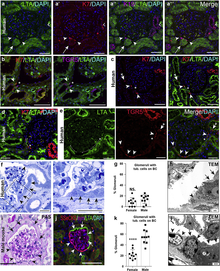
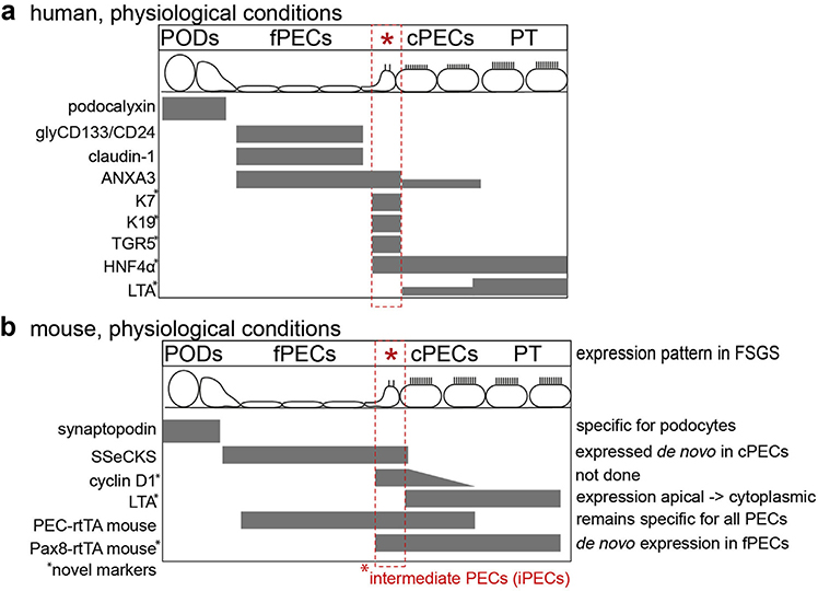
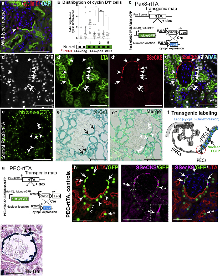
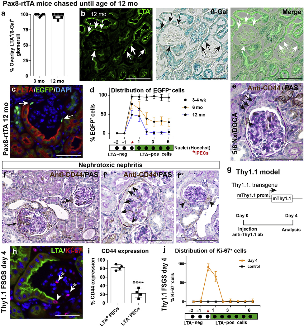
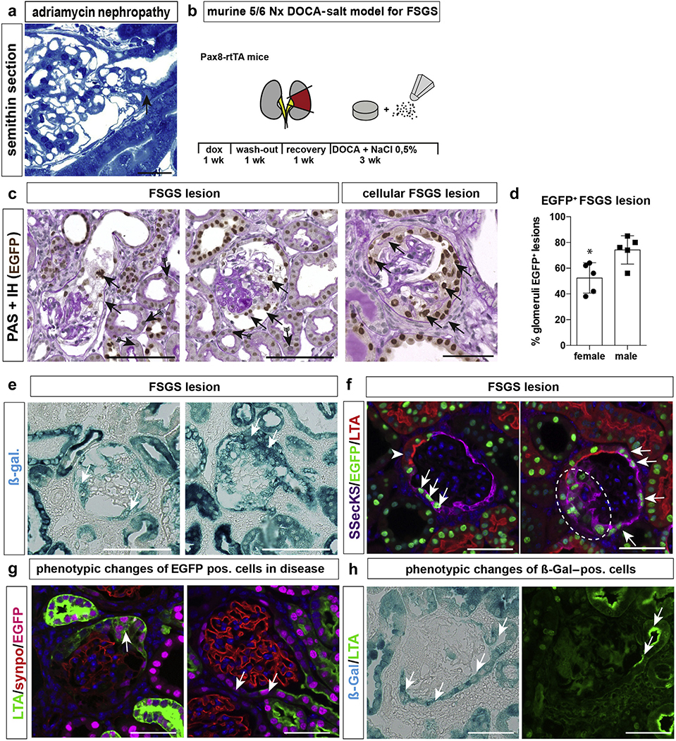
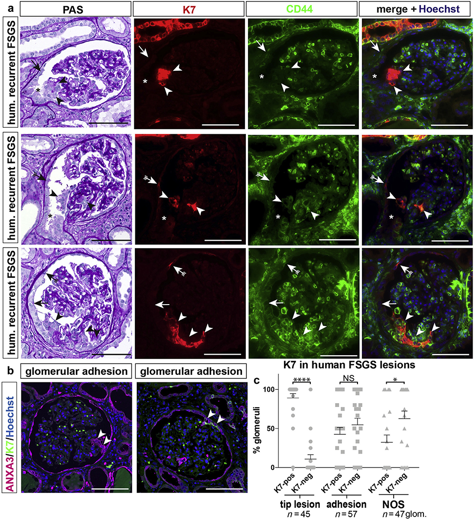
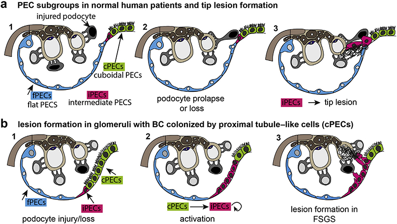
Beside the classical flat parietal epithelial cells (PECs), we investigated proximal tubular epithelial-like cells, a neglected subgroup of PECs. These cells, termed cuboidal PECs, make up the most proximal part of the proximal tubule and may also line parts of Bowman's capsule. Additionally, a third intermediate PEC subgroup was identified at the junction between the flat and cuboidal PEC subgroups at the tubular orifice. The transgenic mouse line PEC-rtTA labeled all three PEC subgroups. Here we show that the inducible Pax8-rtTA mouse line specifically labeled only cuboidal and intermediate PECs, but not flat PECs. In aging Pax8-rtTA mice, cell fate mapping showed no evidence for significant transdifferentiation from flat PECs to cuboidal or intermediate PECs or vice versa. In murine glomerular disease models of crescentic glomerulonephritis, and focal segmental glomerulosclerosis (FSGS), intermediate PECs became more numerous. These intermediate PECs preferentially expressed activation markers CD44 and Ki-67, suggesting that this subgroup of PECs was activated more easily than the classical flat PECs. In mice with FSGS, cuboidal and intermediate PECs formed sclerotic lesions. In patients with FSGS, cells forming the tip lesions expressed markers of intermediate PECs. These novel PEC subgroups form sclerotic lesions and were more prone to cellular activation compared to the classical flat PECs in disease. Thus, colonization of Bowman's capsule by cuboidal PECs may predispose to lesion formation and chronic kidney disease. We propose that tip lesions originate from this novel subgroup of PECs in patients with FSGS.
除了经典的扁平壁上皮细胞 (PECs),我们还研究了近端肾小管上皮样细胞,这是 PECs 中被忽视的亚群。这些细胞被称为立方上皮细胞,构成了近端小管的最近端部分,也可能衬在鲍曼囊的部分区域。此外,在扁平 PEC 亚群和立方 PEC 亚群的管腔开口交界处还鉴定出第三个中间 PEC 亚群。转基因小鼠系 PEC-rtTA 标记了所有三个 PEC 亚群。在这里,我们表明,诱导型 Pax8-rtTA 小鼠系特异性标记仅立方和中间 PECs,但不标记扁平 PECs。在衰老的 Pax8-rtTA 小鼠中,细胞命运图谱显示没有证据表明扁平 PECs 向立方或中间 PECs或反之发生显著的转分化。在新月体性肾小球肾炎和局灶节段性肾小球硬化症 (FSGS) 的小鼠肾小球疾病模型中,中间 PECs 的数量增多。这些中间 PECs 更频繁地表达激活标志物 CD44 和 Ki-67,表明该 PEC 亚群比经典的扁平 PECs 更容易被激活。在 FSGS 小鼠中,立方和中间 PECs 形成硬化病变。在 FSGS 患者中,形成尖端病变的细胞表达中间 PECs 的标志物。与疾病中的经典扁平 PECs 相比,这些新型 PEC 亚群形成硬化病变且更容易发生细胞激活。因此,立方上皮细胞在鲍曼囊的定植可能导致病变形成和慢性肾病。我们提出 FSGS 患者中尖端病变起源于这种新型 PEC 亚群。