• 文献检索
  • 文档翻译
  • 深度研究
  • 学术资讯
  • Suppr Zotero 插件Zotero 插件
  • 邀请有礼
  • 套餐&价格
  • 历史记录
应用&插件
Suppr Zotero 插件Zotero 插件浏览器插件Mac 客户端Windows 客户端微信小程序
定价
高级版会员购买积分包购买API积分包
服务
文献检索文档翻译深度研究API 文档MCP 服务
关于我们
关于 Suppr公司介绍联系我们用户协议隐私条款
关注我们

Suppr 超能文献

核心技术专利:CN118964589B侵权必究
粤ICP备2023148730 号-1Suppr @ 2026

文献检索

告别复杂PubMed语法,用中文像聊天一样搜索,搜遍4000万医学文献。AI智能推荐,让科研检索更轻松。

立即免费搜索

文件翻译

保留排版,准确专业,支持PDF/Word/PPT等文件格式,支持 12+语言互译。

免费翻译文档

深度研究

AI帮你快速写综述,25分钟生成高质量综述,智能提取关键信息,辅助科研写作。

立即免费体验

乳腺非典型增生癌前病变的分子指纹图谱

Molecular fingerprint of precancerous lesions in breast atypical hyperplasia.

作者信息

Zheng Chao, Jia Hong Ying, Liu Li Yuan, Wang Qi, Jiang Hong Chuan, Teng Li Song, Geng Cui Zhi, Jin Feng, Tang Li Li, Zhang Jian Guo, Wang Xiang, Wang Shu, Alejandro Fernandez-Escobar, Wang Fei, Yu Li Xiang, Zhou Fei, Xiang Yu Juan, Huang Shu Ya, Fu Qin Ye, Zhang Qiang, Gao De Zong, Ma Zhong Bing, Li Liang, Fan Zhi Min, Yu Zhi Gang

机构信息

Department of Breast Surgery, The Second Hospital, Cheeloo College of Medicine, Shandong University, Jinan, Shandong, China.

Center of Evidence-based Medicine, The Second Hospital, Cheeloo College of Medicine, Shandong University, Jinan, Shandong, China.

出版信息

J Int Med Res. 2020 Jun;48(6):300060520931616. doi: 10.1177/0300060520931616.

DOI:10.1177/0300060520931616
PMID:32589079
原文链接:https://pmc.ncbi.nlm.nih.gov/articles/PMC7325464/
Abstract

OBJECTIVE

To identify atypical hyperplasia (AH) of the breast by shell-isolated nanoparticle-enhanced Raman spectroscopy (SHINERS), and to explore the molecular fingerprinting characteristics of breast AH.

METHODS

Breast hyperplasia was studied in 11 hospitals across China from January 2015 to December 2016. All patients completed questionnaires on women's health. The differences between patients with and without breast AH were compared. AH breast lesions were detected by Raman spectroscopy followed by the SHINERS technique.

RESULTS

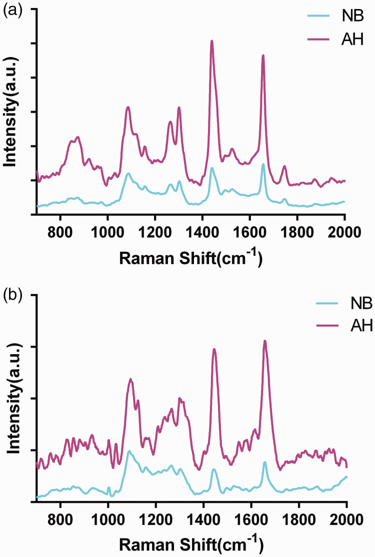
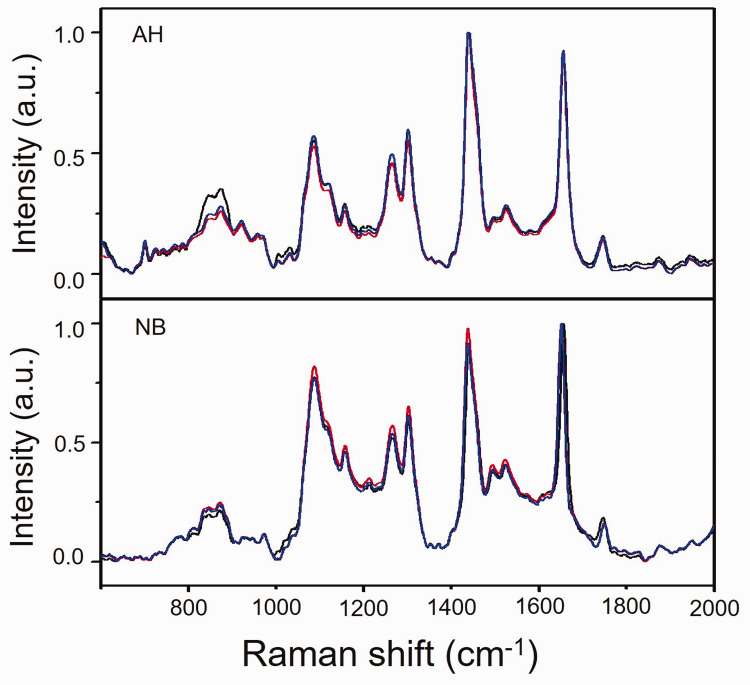
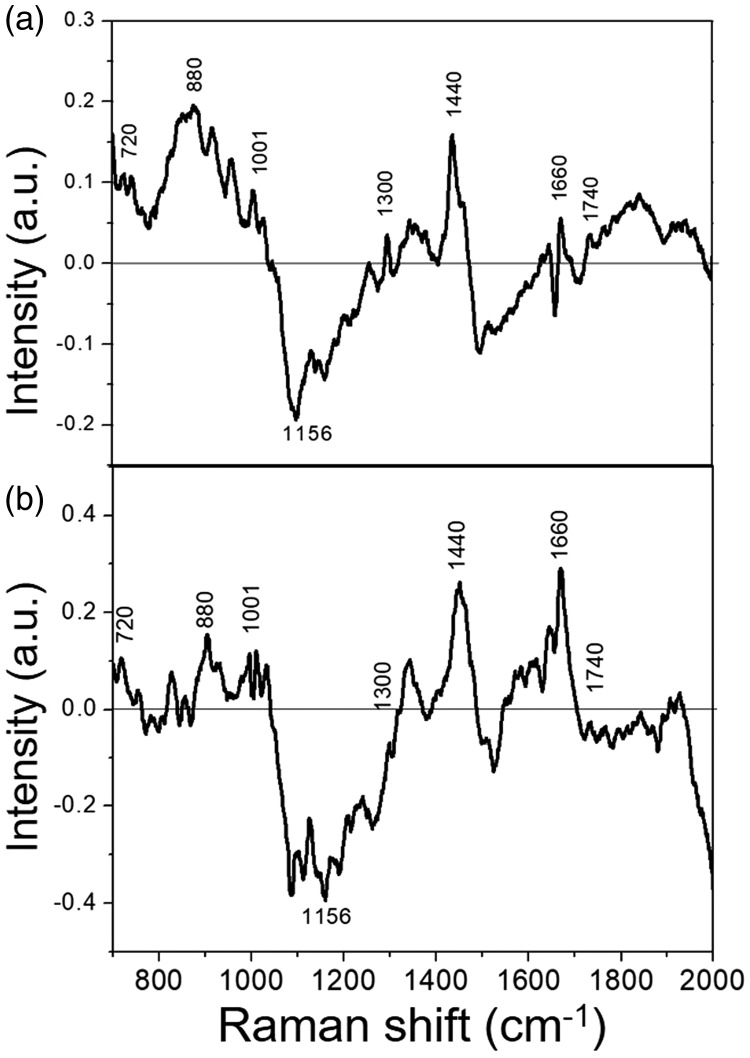
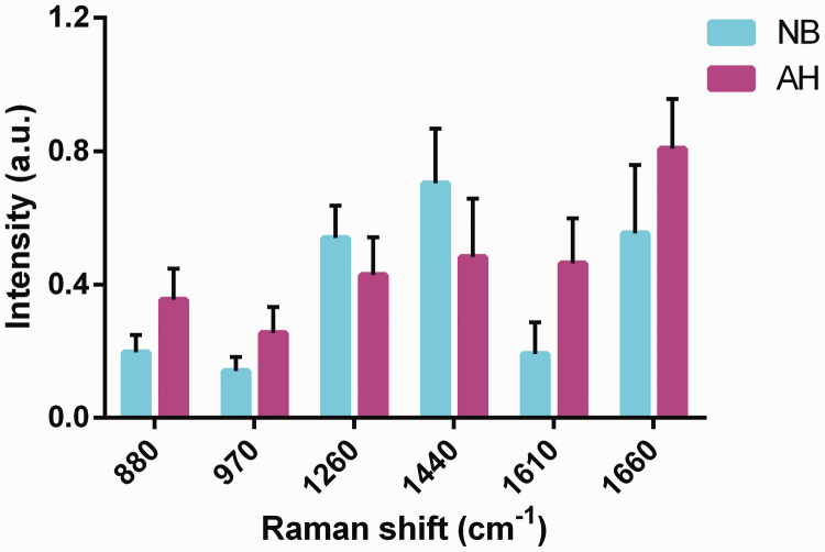
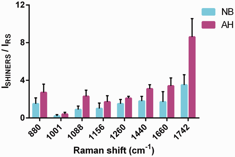
There were no significant differences in clinical features and risk-related factors between patients with breast AH (n = 37) and the control group (n = 2576). Fifteen cases of breast AH lesions were detected by Raman spectroscopy. The main different Raman peaks in patients with AH appeared at 880, 1001, 1086, 1156, 1260, and 1610 cm, attributed to the different vibrational modes of nucleic acids, β-carotene, and proteins. Shell-isolated nanoparticles had different enhancement effects on the nucleic acid, protein, and lipid components in AH.

CONCLUSION

Raman spectroscopy can detect characteristic molecular changes in breast AH lesions, and may thus be useful for the non-invasive early diagnosis and for investigating the mechanism of tumorigenesis in patients with breast AH.

摘要

目的

采用壳层隔离纳米粒子增强拉曼光谱法(SHINERS)鉴别乳腺不典型增生(AH),并探索乳腺AH的分子指纹特征。

方法

2015年1月至2016年12月,在中国11家医院对乳腺增生进行研究。所有患者均完成了关于女性健康的问卷调查。比较有和没有乳腺AH患者之间的差异。通过拉曼光谱法检测AH乳腺病变,随后采用SHINERS技术。

结果

乳腺AH患者(n = 37)与对照组(n = 2576)在临床特征和风险相关因素方面无显著差异。通过拉曼光谱法检测到15例乳腺AH病变。AH患者主要不同的拉曼峰出现在880、1001、1086、1156、1260和1610 cm处,归因于核酸、β-胡萝卜素和蛋白质的不同振动模式。壳层隔离纳米粒子对AH中的核酸、蛋白质和脂质成分具有不同的增强作用。

结论

拉曼光谱法可检测乳腺AH病变中的特征性分子变化,因此可能有助于乳腺AH患者的非侵入性早期诊断及肿瘤发生机制的研究。

https://cdn.ncbi.nlm.nih.gov/pmc/blobs/645b/7325464/e16646e4a4f8/10.1177_0300060520931616-fig5.jpg
https://cdn.ncbi.nlm.nih.gov/pmc/blobs/645b/7325464/3534a0668222/10.1177_0300060520931616-fig1.jpg
https://cdn.ncbi.nlm.nih.gov/pmc/blobs/645b/7325464/e876f30c6117/10.1177_0300060520931616-fig2.jpg
https://cdn.ncbi.nlm.nih.gov/pmc/blobs/645b/7325464/65d60ef5e0c2/10.1177_0300060520931616-fig3.jpg
https://cdn.ncbi.nlm.nih.gov/pmc/blobs/645b/7325464/0f690bb9d841/10.1177_0300060520931616-fig4.jpg
https://cdn.ncbi.nlm.nih.gov/pmc/blobs/645b/7325464/e16646e4a4f8/10.1177_0300060520931616-fig5.jpg

相似文献

1
Molecular fingerprint of precancerous lesions in breast atypical hyperplasia.乳腺非典型增生癌前病变的分子指纹图谱
J Int Med Res. 2020 Jun;48(6):300060520931616. doi: 10.1177/0300060520931616.
2
Exploring type II microcalcifications in benign and premalignant breast lesions by shell-isolated nanoparticle-enhanced Raman spectroscopy (SHINERS).通过壳层隔离纳米粒子增强拉曼光谱(SHINERS)探索乳腺良性和癌前病变中的II型微钙化。
Spectrochim Acta A Mol Biomol Spectrosc. 2014 Nov 11;132:397-402. doi: 10.1016/j.saa.2014.04.147. Epub 2014 May 5.
3
The use of Au@SiO2 shell-isolated nanoparticle-enhanced Raman spectroscopy for human breast cancer detection.金@二氧化硅壳层隔离纳米粒子增强拉曼光谱在人类乳腺癌检测中的应用。
Anal Bioanal Chem. 2014 Sep;406(22):5425-32. doi: 10.1007/s00216-014-7967-5. Epub 2014 Jun 24.
4
Extent of atypical hyperplasia stratifies breast cancer risk in 2 independent cohorts of women.非典型增生程度在两组独立女性队列中对乳腺癌风险进行分层。
Cancer. 2016 Oct;122(19):2971-8. doi: 10.1002/cncr.30153. Epub 2016 Jun 28.
5
Mammographic breast density and risk of breast cancer in women with atypical hyperplasia: an observational cohort study from the Mayo Clinic Benign Breast Disease (BBD) cohort.非典型增生女性的乳腺钼靶密度与乳腺癌风险:一项来自梅奥诊所良性乳腺疾病(BBD)队列的观察性队列研究。
BMC Cancer. 2017 Jan 31;17(1):84. doi: 10.1186/s12885-017-3082-2.
6
The presence of proliferative breast disease with atypia does not significantly influence outcome in early-stage invasive breast cancer treated with conservative surgery and radiation.伴有非典型性的增生性乳腺疾病的存在,对接受保乳手术和放疗的早期浸润性乳腺癌的预后没有显著影响。
Int J Radiat Oncol Biol Phys. 1998 Aug 1;42(1):105-15. doi: 10.1016/s0360-3016(98)00181-3.
7
Microsatellite alterations indicating monoclonality in atypical hyperplasias associated with breast cancer.微卫星改变表明与乳腺癌相关的非典型增生中的单克隆性。
Hum Pathol. 1997 Feb;28(2):214-9. doi: 10.1016/s0046-8177(97)90109-x.
8
Pursuing shell-isolated nanoparticle-enhanced Raman spectroscopy (SHINERS) for concomitant detection of breast lesions and microcalcifications.采用壳层隔离纳米粒子增强拉曼光谱法(SHINERS)同时检测乳腺病变和微钙化。
Nanoscale. 2015 Oct 28;7(40):16960-8. doi: 10.1039/c5nr05319f.
9
Interphase cytogenetics in mammographically detected breast lesions.乳腺钼靶检测到的乳腺病变的间期细胞遗传学
Hum Pathol. 1996 Apr;27(4):330-5. doi: 10.1016/s0046-8177(96)90104-5.
10
Detection of monoclonal microsatellite alterations in atypical breast hyperplasia.非典型乳腺增生中单克隆微卫星改变的检测
J Clin Invest. 1996 Sep 1;98(5):1095-100. doi: 10.1172/JCI118890.

引用本文的文献

1
Raman Spectroscopy of Carotenoid Compounds for Clinical Applications-A Review.用于临床应用的类胡萝卜素化合物的拉曼光谱学研究综述。
Molecules. 2022 Dec 17;27(24):9017. doi: 10.3390/molecules27249017.
2
Diagnosis accuracy of Raman spectroscopy in the diagnosis of breast cancer: a meta-analysis.拉曼光谱在乳腺癌诊断中的诊断准确性:一项荟萃分析。
Anal Bioanal Chem. 2022 Nov;414(27):7911-7922. doi: 10.1007/s00216-022-04326-7. Epub 2022 Sep 23.
3
Diagnostic test of dynamic computed tomography in early gastrointestinal lymphoma and precancerous lesions.

本文引用的文献

1
Atypical ductal hyperplasia: update on diagnosis, management, and molecular landscape.不典型导管增生:诊断、管理和分子特征的最新进展。
Breast Cancer Res. 2018 May 2;20(1):39. doi: 10.1186/s13058-018-0967-1.
2
Label-free Raman spectroscopy provides early determination and precise localization of breast cancer-colonized bone alterations.无标记拉曼光谱可对乳腺癌定植骨改变进行早期测定和精确定位。
Chem Sci. 2017 Nov 15;9(3):743-753. doi: 10.1039/c7sc02905e. eCollection 2018 Jan 21.
3
Molecular profile of atypical hyperplasia of the breast.
动态计算机断层扫描在早期胃肠道淋巴瘤及癌前病变中的诊断试验
Transl Cancer Res. 2022 Jun;11(6):1689-1696. doi: 10.21037/tcr-22-1085.
乳腺非典型性增生的分子特征。
Breast Cancer Res Treat. 2018 Jan;167(1):9-29. doi: 10.1007/s10549-017-4488-x. Epub 2017 Sep 14.
4
Raman spectroscopy for cancer detection and cancer surgery guidance: translation to the clinics.拉曼光谱学在癌症检测和癌症手术指导中的应用:向临床的转化。
Analyst. 2017 Aug 21;142(17):3025-3047. doi: 10.1039/c7an00957g.
5
Biomedical optical spectroscopy for the early diagnosis of gastrointestinal neoplasms.用于胃肠道肿瘤早期诊断的生物医学光学光谱学
Tumour Biol. 2017 Jul;39(7):1010428317717984. doi: 10.1177/1010428317717984.
6
Raman spectroscopy using plasmonic and carbon-based nanoparticles for cancer detection, diagnosis, and treatment guidance.Part 1: Diagnosis.利用等离子体和碳基纳米粒子的拉曼光谱用于癌症检测、诊断和治疗指导。第1部分:诊断。
Drug Metab Rev. 2017 May;49(2):212-252. doi: 10.1080/03602532.2017.1302465. Epub 2017 Jun 15.
7
Label-Free Raman Spectroscopy Detects Stromal Adaptations in Premetastatic Lungs Primed by Breast Cancer.无标记拉曼光谱法检测由乳腺癌引发的肺前转移灶中的基质适应性变化。
Cancer Res. 2017 Jan 15;77(2):247-256. doi: 10.1158/0008-5472.CAN-16-1862. Epub 2016 Nov 15.
8
Pathologic High-risk Lesions, Diagnosis and Management.病理性高危病变:诊断与管理
Clin Obstet Gynecol. 2016 Dec;59(4):727-732. doi: 10.1097/GRF.0000000000000234.
9
Shell-isolated nanoparticle-enhanced Raman spectroscopy study of the adsorption behaviour of DNA bases on Au(111) electrode surfaces.壳层隔绝纳米粒子增强拉曼光谱研究 DNA 碱基在金(111)电极表面的吸附行为。
Analyst. 2016 Jun 21;141(12):3731-6. doi: 10.1039/c6an00180g. Epub 2016 Mar 22.
10
Pursuing shell-isolated nanoparticle-enhanced Raman spectroscopy (SHINERS) for concomitant detection of breast lesions and microcalcifications.采用壳层隔离纳米粒子增强拉曼光谱法(SHINERS)同时检测乳腺病变和微钙化。
Nanoscale. 2015 Oct 28;7(40):16960-8. doi: 10.1039/c5nr05319f.