• 文献检索
  • 文档翻译
  • 深度研究
  • 学术资讯
  • Suppr Zotero 插件Zotero 插件
  • 邀请有礼
  • 套餐&价格
  • 历史记录
应用&插件
Suppr Zotero 插件Zotero 插件浏览器插件Mac 客户端Windows 客户端微信小程序
定价
高级版会员购买积分包购买API积分包
服务
文献检索文档翻译深度研究API 文档MCP 服务
关于我们
关于 Suppr公司介绍联系我们用户协议隐私条款
关注我们

Suppr 超能文献

核心技术专利:CN118964589B侵权必究
粤ICP备2023148730 号-1Suppr @ 2026

文献检索

告别复杂PubMed语法,用中文像聊天一样搜索,搜遍4000万医学文献。AI智能推荐,让科研检索更轻松。

立即免费搜索

文件翻译

保留排版,准确专业,支持PDF/Word/PPT等文件格式,支持 12+语言互译。

免费翻译文档

深度研究

AI帮你快速写综述,25分钟生成高质量综述,智能提取关键信息,辅助科研写作。

立即免费体验

承重时软骨下的压力和灌注。

Subchondral pressures and perfusion during weight bearing.

机构信息

Nuffield Department of Orthopaedics, Rheumatology & Musculoskeletal Sciences, University of Oxford, Botnar Research Centre, Nuffield Orthopaedic Centre, Headington, Oxford, OX3 7LD, UK.

出版信息

J Orthop Surg Res. 2020 Jun 29;15(1):239. doi: 10.1186/s13018-020-01754-y.

DOI:10.1186/s13018-020-01754-y
PMID:32600340
原文链接:https://pmc.ncbi.nlm.nih.gov/articles/PMC7325244/
Abstract

BACKGROUND

Joints withstand huge forces, but little is known about subchondral pressures and perfusion during loading. We developed an in vitro calf foot model to explore intraosseous pressure (IOP) and subchondral perfusion during weight bearing.

METHODS

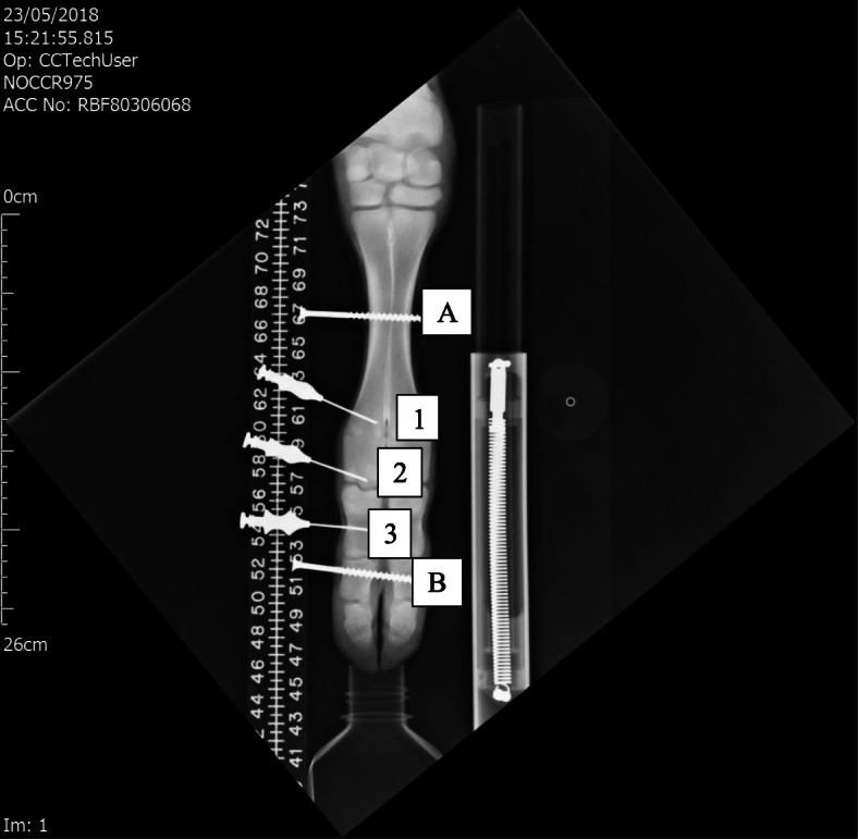
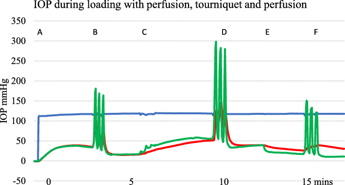
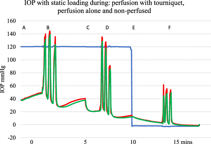
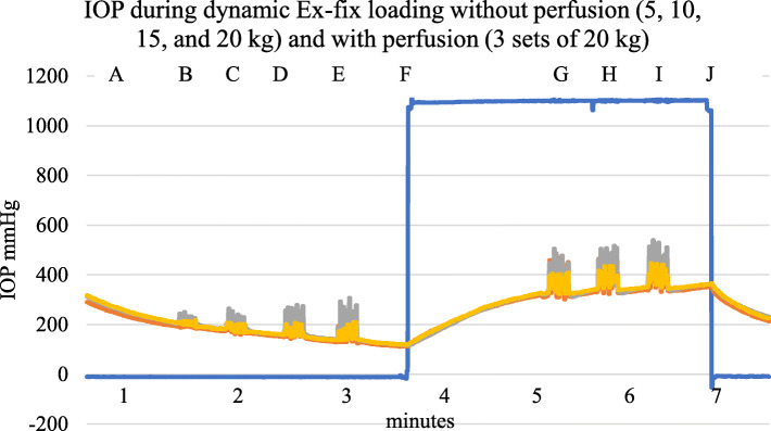
Freshly culled calf forefeet were perfused with serum. IOP was measured at three sites in the foot using intraosseous needles, pressure transducers, and digital recorders. IOP was measured during perfusion, with and without a tourniquet and with differing weights, including static loading and dynamic loading to resemble walking.

RESULTS

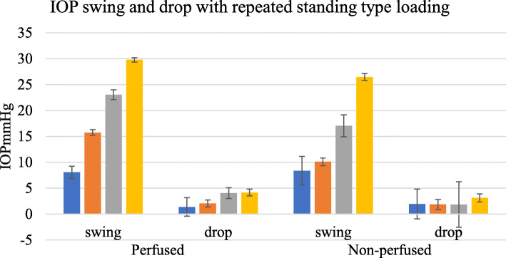
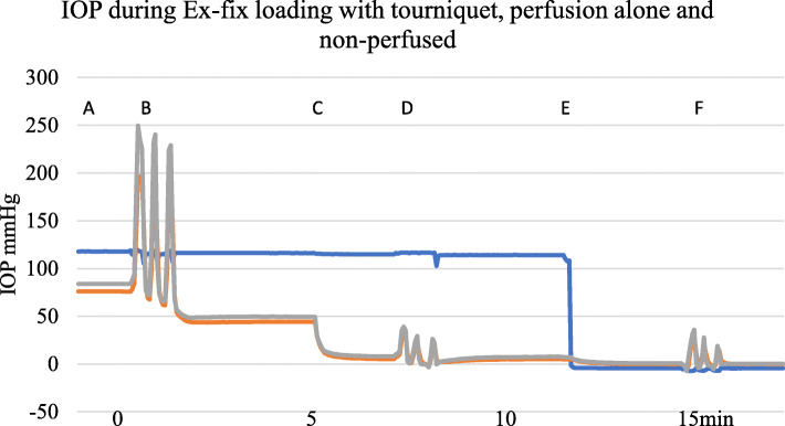
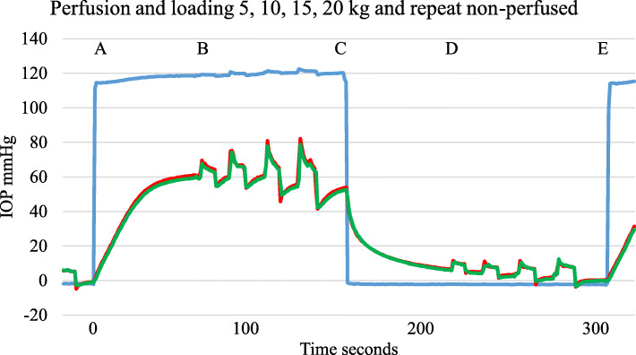
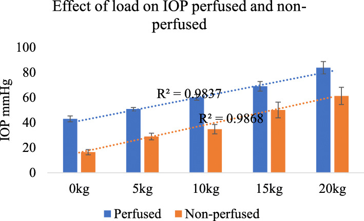
IOP varied with perfusion pressure. Static loading increased subchondral IOP whether the bone was non-perfused, perfused, or perfused with a proximal venous tourniquet (p < 0.0001). Under all perfusion states, IOP was proportional to the load (R = 0.984). Subchondral IOP often exceeded perfusion pressure. On removal of a load, IOP fell to below the pre-load value. Repetitive loading led to a falling IOP whether the foot was perfused or not.

CONCLUSION

Superimposed on a variable background IOP, increased perfusion and physiological loading caused a significant increase in subchondral IOP. Force was thereby transmitted through subchondral bone partly by hydraulic pressure. A falling IOP with repeat loading suggests that there is an intraosseous one-way valve. This offers a new understanding of subchondral perfusion physiology.

摘要

背景

关节承受着巨大的力量,但对于在负重时的软骨下压力和灌注知之甚少。我们开发了一种体外小牛足模型,以探索负重时的骨内压(IOP)和软骨下灌注。

方法

使用血清对新鲜屠宰的小牛前脚进行灌注。使用骨内针、压力传感器和数字记录器在足部的三个部位测量 IOP。在灌注期间、使用和不使用止血带以及不同重量下测量 IOP,包括模拟行走的静态和动态加载。

结果

IOP 随灌注压力而变化。静态加载会增加软骨下 IOP,无论骨骼是否未灌注、灌注或灌注近端静脉止血带(p < 0.0001)。在所有灌注状态下,IOP 与负荷成正比(R = 0.984)。软骨下 IOP 通常超过灌注压力。去除负荷后,IOP 降至低于预加载值。无论足部是否灌注,重复加载都会导致 IOP 下降。

结论

在可变的背景 IOP 上叠加增加的灌注和生理负荷会导致软骨下 IOP 显著增加。因此,力通过软骨下骨部分通过液压传递。重复加载时 IOP 下降表明存在骨内单向阀。这为软骨下灌注生理学提供了新的认识。

https://cdn.ncbi.nlm.nih.gov/pmc/blobs/8995/7325244/2eaec4968913/13018_2020_1754_Fig8_HTML.jpg
https://cdn.ncbi.nlm.nih.gov/pmc/blobs/8995/7325244/381b89a037cb/13018_2020_1754_Fig1_HTML.jpg
https://cdn.ncbi.nlm.nih.gov/pmc/blobs/8995/7325244/fabce01b702e/13018_2020_1754_Fig2_HTML.jpg
https://cdn.ncbi.nlm.nih.gov/pmc/blobs/8995/7325244/8169ed419e05/13018_2020_1754_Fig3_HTML.jpg
https://cdn.ncbi.nlm.nih.gov/pmc/blobs/8995/7325244/88595c7adb96/13018_2020_1754_Fig4_HTML.jpg
https://cdn.ncbi.nlm.nih.gov/pmc/blobs/8995/7325244/e3452e5248c3/13018_2020_1754_Fig5_HTML.jpg
https://cdn.ncbi.nlm.nih.gov/pmc/blobs/8995/7325244/796911043471/13018_2020_1754_Fig6_HTML.jpg
https://cdn.ncbi.nlm.nih.gov/pmc/blobs/8995/7325244/9d6dda39d0b1/13018_2020_1754_Fig7_HTML.jpg
https://cdn.ncbi.nlm.nih.gov/pmc/blobs/8995/7325244/2eaec4968913/13018_2020_1754_Fig8_HTML.jpg

相似文献

1
Subchondral pressures and perfusion during weight bearing.承重时软骨下的压力和灌注。
J Orthop Surg Res. 2020 Jun 29;15(1):239. doi: 10.1186/s13018-020-01754-y.
2
An in vitro model to explore subchondral perfusion and intraosseous pressure.一种用于探索软骨下灌注和骨内压的体外模型。
J Exp Orthop. 2019 Aug 30;6(1):39. doi: 10.1186/s40634-019-0207-3.
3
Intraosseous pressure during loading and with vascular occlusion in an animal model.动物模型中加载及血管闭塞时的骨内压
Bone Joint Res. 2018 Sep 15;7(8):511-516. doi: 10.1302/2046-3758.78.BJR-2017-0343.R2. eCollection 2018 Aug.
4
Factors affecting intraosseous pressure measurement.影响骨内压测量的因素。
J Orthop Surg Res. 2018 Jul 28;13(1):187. doi: 10.1186/s13018-018-0877-z.
5
Walking on water: subchondral vascular physiology explains how joints work and why they become osteoarthritic.水上行走:软骨下血管生理学解释了关节如何运作以及为何会患骨关节炎。
EFORT Open Rev. 2023 Jun 8;8(6):436-442. doi: 10.1530/EOR-23-0002.
6
Hydraulic Joint Function and Osteoarthritis.液压接头的功能与骨关节炎。
JBJS Rev. 2023 Dec 15;11(12). doi: 10.2106/JBJS.RVW.23.00040. eCollection 2023 Dec 1.
7
Marrow fat may distribute the energy of impact loading throughout subchondral bone.骨髓脂肪可能会将冲击载荷的能量分布到软骨下骨中。
Rheumatology (Oxford). 2018 Mar 1;57(3):414-418. doi: 10.1093/rheumatology/kex274.
8
Foot kinematics and loading of professional athletes in American football-specific tasks.美式橄榄球专项任务中职业运动员的足部运动学和受力情况。
Gait Posture. 2013 Sep;38(4):563-9. doi: 10.1016/j.gaitpost.2012.03.034. Epub 2012 May 15.
9
Effect of ankle orientation on heel loading and knee stability for post-stroke individuals wearing ankle-foot orthoses.脚踝方位对中风后佩戴踝足矫形器患者足跟负荷及膝关节稳定性的影响
Prosthet Orthot Int. 2011 Jun;35(2):150-62. doi: 10.1177/0309364611399146. Epub 2011 Mar 29.
10
Intra-articular load distribution in the human ankle joint during motion.人体踝关节运动过程中的关节内负荷分布
Foot Ankle Int. 2001 Mar;22(3):226-33. doi: 10.1177/107110070102200310.

引用本文的文献

1
Walking on water: subchondral vascular physiology explains how joints work and why they become osteoarthritic.水上行走:软骨下血管生理学解释了关节如何运作以及为何会患骨关节炎。
EFORT Open Rev. 2023 Jun 8;8(6):436-442. doi: 10.1530/EOR-23-0002.
2
Subchondral physiology and vasculo-mechanical factors in load transmission and osteoarthritis.软骨下骨生理学及负荷传递与骨关节炎中的血管机械因素
Bone Joint Res. 2021 Sep;10(9):571-573. doi: 10.1302/2046-3758.109.BJR-2021-0031.R1.

本文引用的文献

1
An in vitro model to explore subchondral perfusion and intraosseous pressure.一种用于探索软骨下灌注和骨内压的体外模型。
J Exp Orthop. 2019 Aug 30;6(1):39. doi: 10.1186/s40634-019-0207-3.
2
Intraosseous pressure during loading and with vascular occlusion in an animal model.动物模型中加载及血管闭塞时的骨内压
Bone Joint Res. 2018 Sep 15;7(8):511-516. doi: 10.1302/2046-3758.78.BJR-2017-0343.R2. eCollection 2018 Aug.
3
Factors affecting intraosseous pressure measurement.影响骨内压测量的因素。
J Orthop Surg Res. 2018 Jul 28;13(1):187. doi: 10.1186/s13018-018-0877-z.
4
Marrow fat may distribute the energy of impact loading throughout subchondral bone.骨髓脂肪可能会将冲击载荷的能量分布到软骨下骨中。
Rheumatology (Oxford). 2018 Mar 1;57(3):414-418. doi: 10.1093/rheumatology/kex274.
5
Intraosseous Pressure Monitoring in Healthy Volunteers.健康志愿者的骨内压监测
Prehosp Emerg Care. 2017 Sep-Oct;21(5):567-574. doi: 10.1080/10903127.2017.1302529. Epub 2017 Apr 18.
6
Micro-arteriography.微动脉造影术
Am J Roentgenol Radium Ther. 1948 Jul;60(1):1-12.
7
Could the intraosseous fluid in cancellous bone bear external load significantly within the elastic range?松质骨中的骨内液在弹性范围内能显著承受外部负荷吗?
Proc Inst Mech Eng H. 2004;218(6):375-9. doi: 10.1243/0954411042632153.
8
INTRAOSSEOUS PRESSURE AS A MEASURE OF HEMODYNAMIC CHANGES IN BONE MARROW.骨内压作为衡量骨髓血流动力学变化的指标
Angiology. 1964 Sep;15:396-406. doi: 10.1177/000331976401500903.
9
Hip mechanics.髋关节力学
J Bone Joint Surg Br. 1959 Aug;41-B:550-7. doi: 10.1302/0301-620X.41B3.550.
10
The normal vascular anatomy of the femoral head in adult man.成年男性股骨头的正常血管解剖结构。
J Bone Joint Surg Br. 1953 Aug;35-B(3):442-61. doi: 10.1302/0301-620X.35B3.442.