Harrison Kim D, Hiebert Beverly D, Panahifar Arash, Andronowski Janna M, Ashique Amir M, King Gavin A, Arnason Terra, Swekla Kurtis J, Pivonka Peter, Cooper David Ml
Department of Anatomy, Physiology, and Pharmacology, College of Medicine, University of Saskatchewan, Saskatoon, Canada.
BioMedical Imaging and Therapy Beamline, Canadian Light Source, Saskatoon, Canada.
J Bone Miner Res. 2020 Nov;35(11):2211-2228. doi: 10.1002/jbmr.4124. Epub 2020 Sep 22.
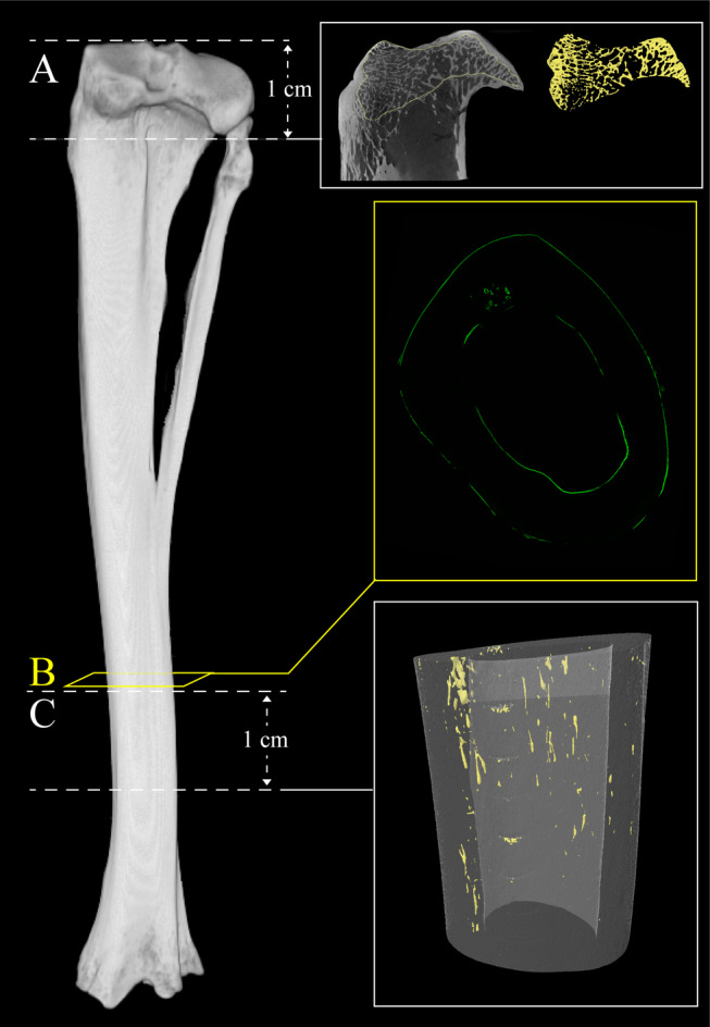
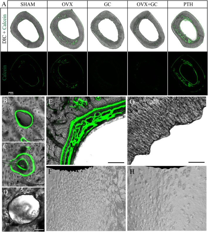
Cortical bone porosity is intimately linked with remodeling, is of growing clinical interest, and is increasingly accessible by imaging. Thus, the potential of animal models of osteoporosis (OP) to provide a platform for studying how porosity develops and responds to interventions is tremendous. To date, rabbit models of OP have largely focused on trabecular microarchitecture or bone density; some such as ovariectomy (OVX) have uncertain efficacy and cortical porosity has not been extensively reported. Our primary objective was to characterize tibial cortical porosity in rabbit-based models of OP, including OVX, glucocorticoids (GC), and OVX + GC relative to controls (SHAM). We sought to: (i) test the hypothesis that intracortical remodeling is elevated in these models; (ii) contrast cortical remodeling and porosity in these models with that induced by parathyroid hormone (1-34; PTH); and (iii) contrast trabecular morphology in the proximal tibia across all groups. Evidence that an increase in cortical porosity occurred in all groups was observed, although this was the least robust for GC. Histomorphometric measures supported the hypothesis that remodeling rate was elevated in all groups and also revealed evidence of uncoupling of bone resorption and formation in the GC and OVX + GC groups. For trabecular bone, a pattern of loss was observed for OVX, GC, and OVX + GC groups, whereas the opposite was observed for PTH. Change in trabecular number best explained these patterns. Taken together, the findings indicated rabbit models provide a viable and varied platform for the study of OP and associated changes in cortical remodeling and porosity. Intriguingly, the evidence revealed differing effects on the cortical and trabecular envelopes for the PTH model. © 2020 The Authors. Journal of Bone and Mineral Research published by Wiley Periodicals LLC on behalf of American Society for Bone and Mineral Research (ASBMR)..
皮质骨孔隙率与骨重塑密切相关,在临床上越来越受到关注,并且通过成像技术越来越容易获取相关信息。因此,骨质疏松症(OP)动物模型为研究孔隙率如何发展以及对干预措施的反应提供平台的潜力巨大。迄今为止,OP兔模型主要集中在小梁微结构或骨密度方面;一些模型,如卵巢切除术(OVX),其疗效尚不确定,且皮质骨孔隙率尚未得到广泛报道。我们的主要目标是在基于兔的OP模型中,包括OVX、糖皮质激素(GC)和OVX + GC模型,相对于对照组(假手术组,SHAM),对胫骨皮质骨孔隙率进行表征。我们试图:(i)检验这些模型中皮质内重塑增加的假设;(ii)将这些模型中的皮质重塑和孔隙率与甲状旁腺激素(1-34;PTH)诱导的情况进行对比;(iii)对比所有组近端胫骨的小梁形态。尽管GC组的情况最不明显,但观察到所有组均出现皮质骨孔隙率增加的证据。组织形态计量学测量结果支持了所有组重塑率均升高的假设,同时也揭示了GC组和OVX + GC组骨吸收与形成解偶联的证据。对于小梁骨,OVX组、GC组和OVX + GC组观察到骨量丢失模式,而PTH组则观察到相反情况。小梁数量的变化最能解释这些模式。综上所述,研究结果表明兔模型为OP以及相关皮质重塑和孔隙率变化的研究提供了一个可行且多样的平台。有趣的是,证据显示PTH模型对皮质骨和小梁骨包膜有不同影响。© 2020作者。《骨与矿物质研究杂志》由Wiley Periodicals LLC代表美国骨与矿物质研究学会(ASBMR)出版。