Pediatric Surgery Division, Department of Surgery, Faculty of Medicine, Public Health and Nursing, Universitas Gadjah Mada/Dr. Sardjito Hospital, Jl. Kesehatan No. 1, Yogyakarta, 55281, Indonesia.
Department of Anatomical Pathology, Faculty of Medicine, Public Health and Nursing, Universitas Gadjah Mada/Dr. Sardjito Hospital, Yogyakarta, 55281, Indonesia.
Diagn Pathol. 2020 Jul 2;15(1):79. doi: 10.1186/s13000-020-00996-y.
Without early recognition and Kasai procedure, biliary atresia (BA) results in liver cirrhosis and leads to either transplantation or death at a young age. We aimed to characterize the liver histopathological findings for prediction of cirrhosis and survival in BA patients after Kasai surgery.
We retrospectively reviewed all histopathological results for BA patients who underwent liver biopsy during Kasai surgery from August 2012 to December 2018 in Dr. Sardjito Hospital, Yogyakarta, Indonesia.
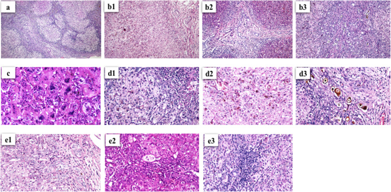
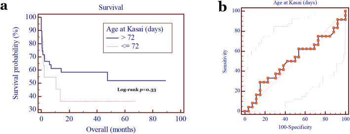
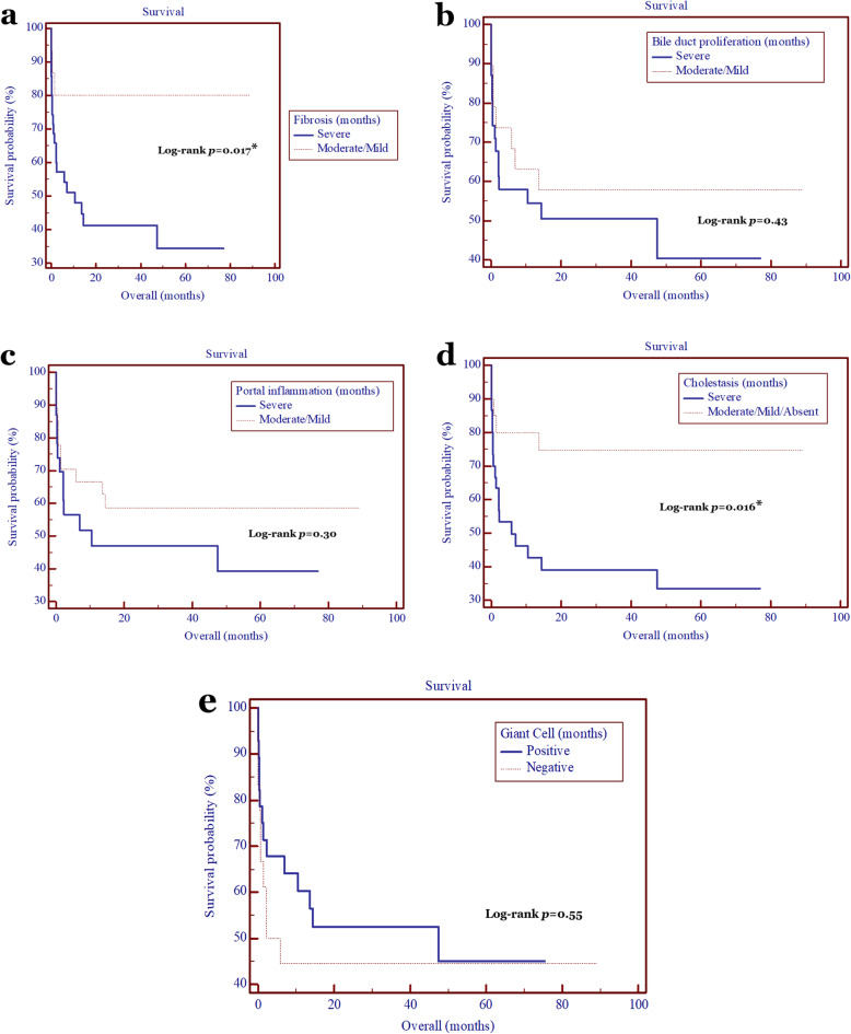
Fifty infants with BA were ascertained in our study, of whom 27 were males and 23 were females. The median age of Kasai procedure was 102.5 days (interquartile range (IQR), 75.75-142.25 days). There were 33 (66%) and 17 (34%) BA patients with and without liver cirrhosis, respectively, while the overall survival was 52%. The patients with a severe bile duct proliferation, severe cholestasis, and severe portal inflammation have a higher risk by 27-, 22-, and 19.3-fold, respectively, to develop liver cirrhosis compared with patients with a moderate/mild bile duct proliferation, moderate/mild/without cholestasis, and moderate/mild portal inflammation, respectively (p = 3.6 × 10, 5.6 × 10, and 1.6 × 10, respectively), while the giant cell transformation was not associate with the development of liver cirrhosis (p = 0.77). The bile duct proliferation was strongly correlated with cholestasis and portal inflammation (p = 7.3 × 10 and 2 × 10, respectively), and cholestasis was also significantly correlated with portal inflammation (p = 0.016). Interestingly, the age at Kasai procedure was strongly associated with the development of liver cirrhosis (p = 0.02), but not with the patients' survival (p = 0.33), while the degree of fibrosis and cholestasis were significantly correlated with the patients' survival, with HR of 3.9 (95% CI = 1.7-9.0; p = 0.017) and 3.1 (95% CI = 1.4-7.0; p = 0.016), respectively.
Histopathological findings of bile duct proliferation, cholestasis, and portal inflammation can predict the liver cirrhosis development in patients with BA. Furthermore, degree of fibrosis and cholestasis affect the patients' survival following the Kasai operation.
如果不能早期识别并进行葛西手术,胆道闭锁(BA)会导致肝硬化,患儿在年幼时就需要进行肝移植或死亡。本研究旨在分析胆道闭锁患儿葛西手术后的肝脏组织病理学特征,预测肝硬化的发生和患儿的生存情况。
我们回顾性分析了 2012 年 8 月至 2018 年 12 月期间在印度尼西亚日惹的萨德尔吉托医院接受葛西手术的所有胆道闭锁患儿的肝活检组织病理学结果。
本研究共纳入了 50 例胆道闭锁患儿,其中男 27 例,女 23 例。葛西手术的中位年龄为 102.5 天(四分位距(IQR):75.75-142.25 天)。33 例(66%)和 17 例(34%)患儿分别发展为肝硬化和无肝硬化,总体生存率为 52%。与中/轻度胆管增生、中/轻度/无胆汁淤积和中/轻度门静脉炎症患儿相比,重度胆管增生、重度胆汁淤积和重度门静脉炎症患儿发生肝硬化的风险分别增加 27 倍、22 倍和 19.3 倍(p=3.6×10、5.6×10 和 1.6×10,分别),而巨细胞转化与肝硬化的发生无关(p=0.77)。胆管增生与胆汁淤积和门静脉炎症显著相关(p=7.3×10 和 2×10,分别),胆汁淤积也与门静脉炎症显著相关(p=0.016)。有趣的是,葛西手术年龄与肝硬化的发生密切相关(p=0.02),但与患儿的生存率无关(p=0.33),而纤维化和胆汁淤积程度与患儿的生存率显著相关,HR 分别为 3.9(95%CI:1.7-9.0;p=0.017)和 3.1(95%CI:1.4-7.0;p=0.016)。
胆管增生、胆汁淤积和门静脉炎症的组织病理学发现可预测胆道闭锁患儿肝硬化的发生。此外,纤维化和胆汁淤积程度影响患儿葛西手术后的生存情况。