• 文献检索
  • 文档翻译
  • 深度研究
  • 学术资讯
  • Suppr Zotero 插件Zotero 插件
  • 邀请有礼
  • 套餐&价格
  • 历史记录
应用&插件
Suppr Zotero 插件Zotero 插件浏览器插件Mac 客户端Windows 客户端微信小程序
定价
高级版会员购买积分包购买API积分包
服务
文献检索文档翻译深度研究API 文档MCP 服务
关于我们
关于 Suppr公司介绍联系我们用户协议隐私条款
关注我们

Suppr 超能文献

核心技术专利:CN118964589B侵权必究
粤ICP备2023148730 号-1Suppr @ 2026

文献检索

告别复杂PubMed语法,用中文像聊天一样搜索,搜遍4000万医学文献。AI智能推荐,让科研检索更轻松。

立即免费搜索

文件翻译

保留排版,准确专业,支持PDF/Word/PPT等文件格式,支持 12+语言互译。

免费翻译文档

深度研究

AI帮你快速写综述,25分钟生成高质量综述,智能提取关键信息,辅助科研写作。

立即免费体验

纳入[F]FDG PET/CT 影像组学的小涎腺癌患者预后模型的开发与验证

Development and validation of a prognostic model incorporating [F]FDG PET/CT radiomics for patients with minor salivary gland carcinoma.

作者信息

Cheng Nai-Ming, Hsieh Cheng-En, Fang Yu-Hua Dean, Liao Chun-Ta, Ng Shu-Hang, Wang Hung-Ming, Chou Wen-Chi, Lin Chien-Yu, Yen Tzu-Chen

机构信息

Department of Nuclear Medicine and Molecular Imaging Center, Chang Gung Memorial Hospital, Chang Gung University College of Medicine, Taoyuan, Taiwan.

Department of Nuclear Medicine, Chang Gung Memorial Hospital, Keelung, Taiwan.

出版信息

EJNMMI Res. 2020 Jul 6;10(1):74. doi: 10.1186/s13550-020-00631-3.

DOI:10.1186/s13550-020-00631-3
PMID:32632638
原文链接:https://pmc.ncbi.nlm.nih.gov/articles/PMC7338312/
Abstract

OBJECTIVES

The aim of this study was to develop and validate a prognostic model incorporating [F]FDG PET/CT radiomics for patients of minor salivary gland carcinoma (MSGC).

METHODS

We retrospectively reviewed the pretreatment [F]FDG PET/CT images of 75 MSGC patients treated with curative intent. Using a 1.5:1 ratio, the patients were randomly divided into a training and validation group. The main outcome measurements were overall survival (OS) and relapse-free survival (RFS). All of the patients were followed up for at least 30 months or until death. Following segmentation of tumors and lymph nodes on PET images, radiomic features were extracted. The prognostic significance of PET radiomics and clinical parameters in the training group was examined using receiver operating characteristic curve analysis. Variables showing a significant impact on OS and RFS were entered into multivariable Cox regression models. Recursive partitioning analysis was subsequently implemented to devise a prognostic index, whose performance was examined in the validation group. Finally, the performance of the index was compared with clinical variables in the entire cohort and nomograms for surgically treated cases.

RESULTS

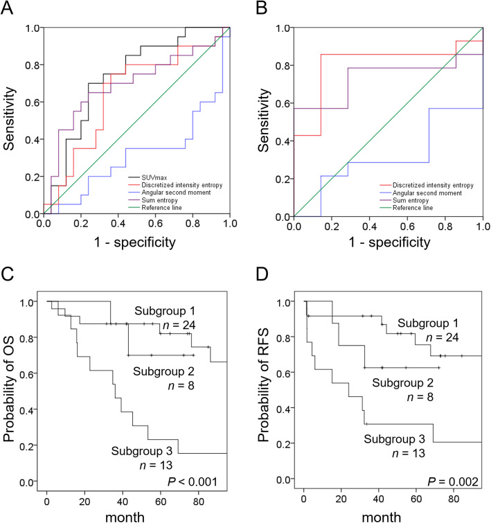
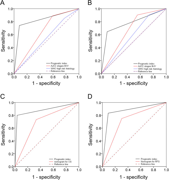
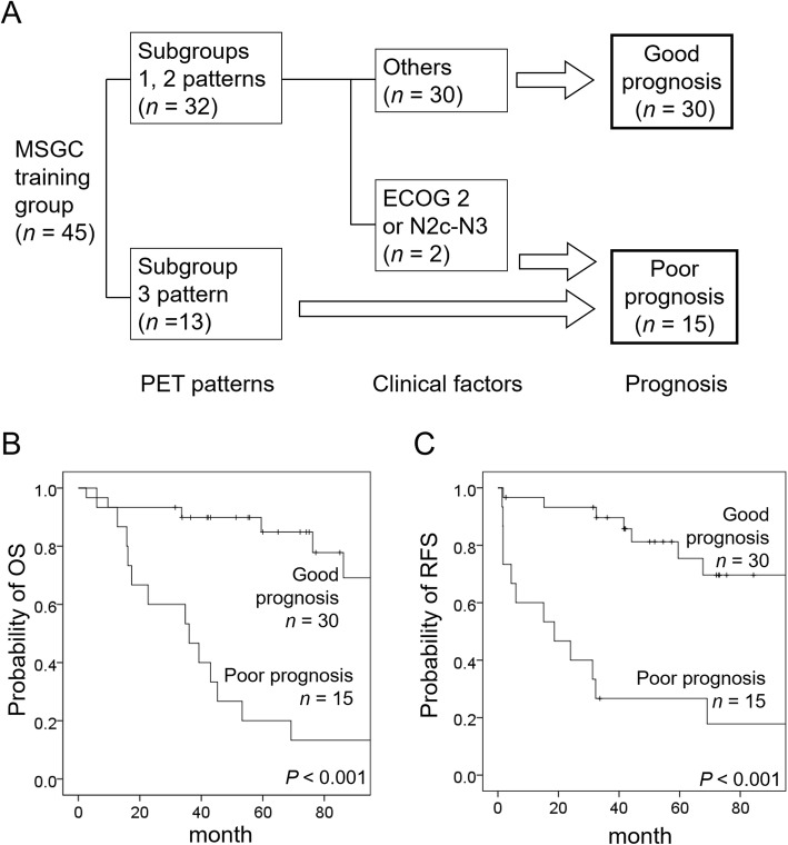
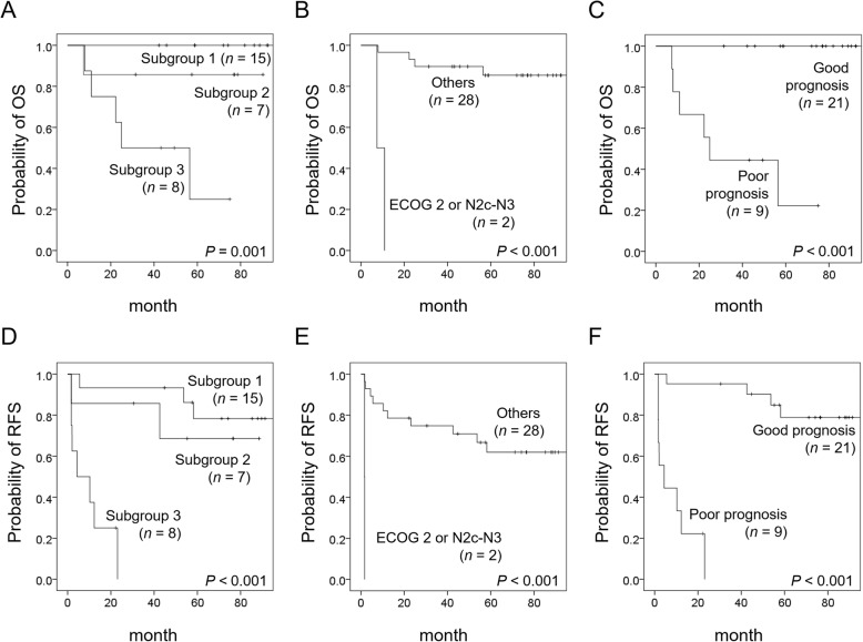
The training and validation groups consisted of 45 and 30 patients, respectively. The median follow-up time in the entire cohort was 59.5 months. Eighteen relapse, 19 dead, and thirteen relapse, eight dead events were found in the training and validation cohorts, respectively. In the training group, two factors were identified as independently associated with poor OS, i.e., (1) tumors with both high maximum standardized uptake value (SUV) and discretized intensity entropy and (2) poor performance status or N2c-N3 stage. A prognostic model based on the above factors was devised and showed significant higher concordance index (C-index) for OS than those of AJCC stage and high-risk histology (C-index: 0.83 vs. 0.65, P = 0.005; 0.83 vs. 0.54, P < 0.001, respectively). This index also demonstrated superior performance than nomogram for OS (C-index: 0.88 vs. 0.70, P = 0.017) and that for RFS (C-index: 0.87 vs. 0.72, P = 0.004).

CONCLUSIONS

We devised a novel prognostic model that incorporates [F]FDG PET/CT radiomics and may help refine outcome prediction in patients with MSGC.

摘要

目的

本研究旨在开发并验证一种纳入[F]FDG PET/CT影像组学的小涎腺癌(MSGC)患者预后模型。

方法

我们回顾性分析了75例接受根治性治疗的MSGC患者的治疗前[F]FDG PET/CT图像。按照1.5:1的比例,将患者随机分为训练组和验证组。主要观察指标为总生存期(OS)和无复发生存期(RFS)。所有患者均随访至少30个月或直至死亡。在PET图像上对肿瘤和淋巴结进行分割后,提取影像组学特征。使用受试者工作特征曲线分析来检验训练组中PET影像组学和临床参数的预后意义。将对OS和RFS有显著影响的变量纳入多变量Cox回归模型。随后进行递归划分分析以设计一个预后指数,并在验证组中检验其性能。最后,在整个队列中将该指数的性能与临床变量进行比较,并为手术治疗病例绘制列线图。

结果

训练组和验证组分别有45例和30例患者。整个队列的中位随访时间为59.5个月。训练组和验证组分别发现18例复发、1... 展开 19例死亡,以及13例复发、8例死亡事件。在训练组中,有两个因素被确定与不良OS独立相关,即:(1)最大标准化摄取值(SUV)高且离散强度熵高的肿瘤,以及(2)身体状况差或N2c - N3期。基于上述因素设计了一个预后模型,该模型显示出比AJCC分期和高危组织学更高的OS一致性指数(C指数)(C指数:0... 展开 83对0.65,P = 0.005;0.83对0.54,P < 0.001)。该指数在OS方面也显示出比列线图更好的性能(C指数: 0.88对0.70,P = 0.017)以及在RFS方面(C指数: 0.87对0.72,P = 0.004)。

结论

我们设计了一种纳入[F]FDG PET/CT影像组学的新型预后模型,可能有助于优化MSGC患者的预后预测。

https://cdn.ncbi.nlm.nih.gov/pmc/blobs/c538/7338312/6b32cc6a90fa/13550_2020_631_Fig4_HTML.jpg
https://cdn.ncbi.nlm.nih.gov/pmc/blobs/c538/7338312/662deaeb6502/13550_2020_631_Fig1_HTML.jpg
https://cdn.ncbi.nlm.nih.gov/pmc/blobs/c538/7338312/8540d49c410a/13550_2020_631_Fig2_HTML.jpg
https://cdn.ncbi.nlm.nih.gov/pmc/blobs/c538/7338312/91d3d6966fbe/13550_2020_631_Fig3_HTML.jpg
https://cdn.ncbi.nlm.nih.gov/pmc/blobs/c538/7338312/6b32cc6a90fa/13550_2020_631_Fig4_HTML.jpg

相似文献

1
Development and validation of a prognostic model incorporating [F]FDG PET/CT radiomics for patients with minor salivary gland carcinoma.纳入[F]FDG PET/CT 影像组学的小涎腺癌患者预后模型的开发与验证
EJNMMI Res. 2020 Jul 6;10(1):74. doi: 10.1186/s13550-020-00631-3.
2
Prognostic Value of Tumor Heterogeneity and SUVmax of Pretreatment 18F-FDG PET/CT for Salivary Gland Carcinoma With High-Risk Histology.高风险组织病理学唾液腺癌中肿瘤异质性和预处理 18F-FDG PET/CT 的 SUVmax 的预后价值。
Clin Nucl Med. 2019 May;44(5):351-358. doi: 10.1097/RLU.0000000000002530.
3
Prognostic value of F-FDG PET radiomics and sarcopenia in patients with oral squamous cell carcinoma.18F-FDG PET 影像组学与骨骼肌减少症对口腔鳞状细胞癌患者预后的预测价值。
Med Phys. 2024 Jul;51(7):4907-4921. doi: 10.1002/mp.16949. Epub 2024 Jan 22.
4
Radiomics Analysis of PET and CT Components of F-FDG PET/CT Imaging for Prediction of Progression-Free Survival in Advanced High-Grade Serous Ovarian Cancer.用于预测晚期高级别浆液性卵巢癌无进展生存期的¹⁸F-FDG PET/CT成像中PET与CT成分的影像组学分析
Front Oncol. 2021 Apr 13;11:638124. doi: 10.3389/fonc.2021.638124. eCollection 2021.
5
Prognostic Value of F-FDG PET/CT Radiomics in Extranodal Nasal-Type NK/T Cell Lymphoma.F-FDG PET/CT 影像组学对结外鼻型 NK/T 细胞淋巴瘤的预后价值
Korean J Radiol. 2024 Feb;25(2):189-198. doi: 10.3348/kjr.2023.0618.
6
Radiomics Analysis of PET and CT Components of PET/CT Imaging Integrated with Clinical Parameters: Application to Prognosis for Nasopharyngeal Carcinoma.PET/CT 影像 PET 成分和 CT 成分的放射组学分析与临床参数相结合:在鼻咽癌预后中的应用。
Mol Imaging Biol. 2019 Oct;21(5):954-964. doi: 10.1007/s11307-018-01304-3.
7
Development and Validation of an F-FDG PET/CT-based Radiomics Nomogram for Predicting the Prognosis of Patients with Esophageal Squamous Cell Carcinoma.基于 F-FDG PET/CT 影像组学构建列线图预测食管鳞癌患者预后
Acad Radiol. 2024 Dec;31(12):5066-5077. doi: 10.1016/j.acra.2024.05.029. Epub 2024 Jun 6.
8
Development and validation of an F-FDG PET radiomic model for prognosis prediction in patients with nasal-type extranodal natural killer/T cell lymphoma.建立并验证 ^18F-FDG PET 影像组学模型预测鼻型结外自然杀伤/T 细胞淋巴瘤患者预后
Eur Radiol. 2020 Oct;30(10):5578-5587. doi: 10.1007/s00330-020-06943-1. Epub 2020 May 20.
9
PET/CT Radiomic Features: A Potential Biomarker for EGFR Mutation Status and Survival Outcome Prediction in NSCLC Patients Treated With TKIs.PET/CT影像组学特征:非小细胞肺癌患者接受酪氨酸激酶抑制剂治疗时表皮生长因子受体突变状态及生存结果预测的潜在生物标志物
Front Oncol. 2022 Jun 21;12:894323. doi: 10.3389/fonc.2022.894323. eCollection 2022.
10
Development and validation of F-FDG PET/CT radiomics-based nomogram to predict visceral pleural invasion in solid lung adenocarcinoma.基于F-FDG PET/CT影像组学的列线图预测实性肺腺癌脏层胸膜侵犯的开发与验证
Ann Nucl Med. 2023 Nov;37(11):605-617. doi: 10.1007/s12149-023-01861-w. Epub 2023 Aug 20.

引用本文的文献

1
Methodological evaluation of original articles on radiomics and machine learning for outcome prediction based on positron emission tomography (PET).基于正电子发射断层扫描(PET)的放射组学和机器学习的预后预测的原始文章的方法学评价。
Nuklearmedizin. 2023 Dec;62(6):361-369. doi: 10.1055/a-2198-0545. Epub 2023 Nov 23.
2
A systematic review and meta-analysis of predictive and prognostic models for outcome prediction using positron emission tomography radiomics in head and neck squamous cell carcinoma patients.头颈部鳞状细胞癌患者使用正电子发射断层扫描放射组学进行预后预测的预测和预后模型的系统评价和荟萃分析。
Cancer Med. 2023 Aug;12(15):16181-16194. doi: 10.1002/cam4.6278. Epub 2023 Jun 24.
3

本文引用的文献

1
The Image Biomarker Standardization Initiative: Standardized Quantitative Radiomics for High-Throughput Image-based Phenotyping.影像生物标志物标准化倡议:高通量基于影像表型的标准化定量放射组学。
Radiology. 2020 May;295(2):328-338. doi: 10.1148/radiol.2020191145. Epub 2020 Mar 10.
2
Radiomics in nuclear medicine: robustness, reproducibility, standardization, and how to avoid data analysis traps and replication crisis.核医学中的放射组学:稳健性、可重复性、标准化,以及如何避免数据分析陷阱和再现性危机。
Eur J Nucl Med Mol Imaging. 2019 Dec;46(13):2638-2655. doi: 10.1007/s00259-019-04391-8. Epub 2019 Jun 25.
3
Associations between GLUT expression and SUV values derived from FDG-PET in different tumors-A systematic review and meta analysis.
The added values of F-FDG PET/CT in differentiating cancer recurrence and osteoradionecrosis of mandible in patients with treated oral squamous cell carcinoma.
F-FDG PET/CT在鉴别经治疗的口腔鳞状细胞癌患者下颌骨癌复发与放射性骨坏死中的附加价值。
EJNMMI Res. 2023 Apr 3;13(1):25. doi: 10.1186/s13550-023-00965-8.
4
The Role of Radiomics in Salivary Gland Imaging: A Systematic Review and Radiomics Quality Assessment.放射组学在唾液腺成像中的作用:系统评价与放射组学质量评估
Diagnostics (Basel). 2022 Dec 1;12(12):3002. doi: 10.3390/diagnostics12123002.
5
Radiomics in Oncological PET Imaging: A Systematic Review-Part 1, Supradiaphragmatic Cancers.肿瘤PET成像中的放射组学:系统综述 - 第1部分,膈上癌症
Diagnostics (Basel). 2022 May 27;12(6):1329. doi: 10.3390/diagnostics12061329.
6
Differentiating low and high grade mucoepidermoid carcinoma of the salivary glands using CT radiomics.利用CT影像组学鉴别涎腺低级别和高级别黏液表皮样癌
Gland Surg. 2021 May;10(5):1646-1654. doi: 10.21037/gs-20-830.
7
A Systematic Review of PET Textural Analysis and Radiomics in Cancer.正电子发射断层扫描(PET)纹理分析与放射组学在癌症中的系统评价
Diagnostics (Basel). 2021 Feb 23;11(2):380. doi: 10.3390/diagnostics11020380.
8
Weakly supervised deep learning for determining the prognostic value of F-FDG PET/CT in extranodal natural killer/T cell lymphoma, nasal type.基于弱监督学习的 F-FDG PET/CT 预测结外鼻型自然杀伤/T 细胞淋巴瘤预后的价值研究。
Eur J Nucl Med Mol Imaging. 2021 Sep;48(10):3151-3161. doi: 10.1007/s00259-021-05232-3. Epub 2021 Feb 20.
不同肿瘤中葡萄糖转运蛋白表达与 FDG-PET 摄取值 SUV 的相关性:系统评价和荟萃分析。
PLoS One. 2019 Jun 17;14(6):e0217781. doi: 10.1371/journal.pone.0217781. eCollection 2019.
4
Minor salivary gland tumors of the head and neck-Memorial Sloan Kettering experience: Incidence and outcomes by site and histological type.头颈部小唾液腺肿瘤-纪念斯隆凯特琳经验:部位和组织学类型的发生率和结果。
Cancer. 2019 Oct 1;125(19):3354-3366. doi: 10.1002/cncr.32208. Epub 2019 Jun 7.
5
Prognostic Value of Tumor Heterogeneity and SUVmax of Pretreatment 18F-FDG PET/CT for Salivary Gland Carcinoma With High-Risk Histology.高风险组织病理学唾液腺癌中肿瘤异质性和预处理 18F-FDG PET/CT 的 SUVmax 的预后价值。
Clin Nucl Med. 2019 May;44(5):351-358. doi: 10.1097/RLU.0000000000002530.
6
Minimum sample size for developing a multivariable prediction model: PART II - binary and time-to-event outcomes.建立多变量预测模型的最小样本量:第二部分 - 二分类和生存数据。
Stat Med. 2019 Mar 30;38(7):1276-1296. doi: 10.1002/sim.7992. Epub 2018 Oct 24.
7
Pretreatment Primary Tumor and Nodal SUVmax Values on 18F-FDG PET/CT Images Predict Prognosis in Patients With Salivary Gland Carcinoma.18F-FDG PET/CT 图像上原发肿瘤和淋巴结 SUVmax 值预测唾液腺癌患者的预后。
Clin Nucl Med. 2018 Dec;43(12):869-879. doi: 10.1097/RLU.0000000000002287.
8
Correlations between metabolic texture features, genetic heterogeneity, and mutation burden in patients with lung cancer.肺癌患者代谢纹理特征、遗传异质性和突变负担之间的相关性。
Eur J Nucl Med Mol Imaging. 2019 Feb;46(2):446-454. doi: 10.1007/s00259-018-4138-5. Epub 2018 Aug 25.
9
Development of a novel salivary gland cancer lymph node staging system.一种新型唾液腺癌淋巴结分期系统的建立。
Cancer. 2018 Aug 1;124(15):3171-3180. doi: 10.1002/cncr.31535. Epub 2018 May 9.
10
Heterogeneity and irregularity of pretreatment F-fluorodeoxyglucose positron emission tomography improved prognostic stratification of p16-negative high-risk squamous cell carcinoma of the oropharynx.预处理 F-氟代脱氧葡萄糖正电子发射断层扫描的异质性和不规则性改善了 p16 阴性高危口咽鳞癌的预后分层。
Oral Oncol. 2018 Mar;78:156-162. doi: 10.1016/j.oraloncology.2018.01.030. Epub 2018 Feb 20.