• 文献检索
  • 文档翻译
  • 深度研究
  • 学术资讯
  • Suppr Zotero 插件Zotero 插件
  • 邀请有礼
  • 套餐&价格
  • 历史记录
应用&插件
Suppr Zotero 插件Zotero 插件浏览器插件Mac 客户端Windows 客户端微信小程序
定价
高级版会员购买积分包购买API积分包
服务
文献检索文档翻译深度研究API 文档MCP 服务
关于我们
关于 Suppr公司介绍联系我们用户协议隐私条款
关注我们

Suppr 超能文献

核心技术专利:CN118964589B侵权必究
粤ICP备2023148730 号-1Suppr @ 2026

文献检索

告别复杂PubMed语法,用中文像聊天一样搜索,搜遍4000万医学文献。AI智能推荐,让科研检索更轻松。

立即免费搜索

文件翻译

保留排版,准确专业,支持PDF/Word/PPT等文件格式,支持 12+语言互译。

免费翻译文档

深度研究

AI帮你快速写综述,25分钟生成高质量综述,智能提取关键信息,辅助科研写作。

立即免费体验

PET/CT联合胸部增强CT在评估成年患者原发性心脏肿瘤中的应用

Combined PET/CT with thoracic contrast-enhanced CT in assessment of primary cardiac tumors in adult patients.

作者信息

Liu En-Tao, Sun Tao-Tao, Dong Hao-Jian, Wang Si-Yun, Chen Ze-Rui, Liu Chao, Shao Dan, Lian Zhou-Yang, Xie Qiu, Wang Shu-Xia

机构信息

WeiLun PET Center, Department of Nuclear Medicine, Guangdong Provincial People's Hospital, Guangdong Academy of Medical Sciences, Room 517, 5/F, Weilun Building of Guangdong Provincial People's Hospital, 106 Zhongshan Er Road, Guangzhou, 510080, Guangdong, People's Republic of China.

Department of Cardiology, Guangdong Provincial People's Hospital, Guangdong Academy of Medical Sciences, 106 Zhongshan Er Road, Guangzhou, 510080, Guangdong, People's Republic of China.

出版信息

EJNMMI Res. 2020 Jul 6;10(1):75. doi: 10.1186/s13550-020-00661-x.

DOI:10.1186/s13550-020-00661-x
PMID:32632639
原文链接:https://pmc.ncbi.nlm.nih.gov/articles/PMC7338301/
Abstract

BACKGROUND

F-FDG PET/CT is a key molecular imaging modality to noninvasively assess and differentiate benign and malignant cardiac tumors. However, few benign cardiac tumors can be characterized by increased F-FDG uptake, which makes differential diagnosis difficult. This study sought to retrospectively evaluate whether combined F-FDG PET/CT with thoracic contrast-enhanced CT (CECT) helps in assessing primary cardiac tumors in adult patients, compared with CECT or PET/CT alone.

METHODS

Forty-six consecutive patients who were diagnosed as primary cardiac tumors were enrolled. All patients underwent F-FDG PET/CT followed by thoracic CECT before biopsy or surgery. Visual qualitative interpretation and quantitative analysis were performed, and diagnostic performance was evaluated.

RESULTS

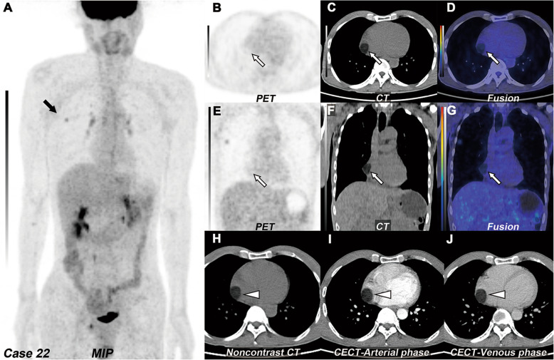
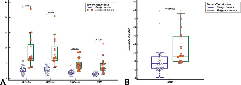
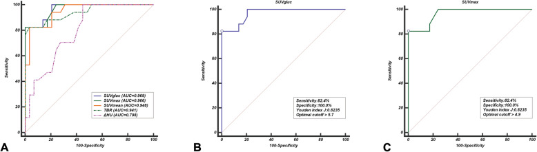
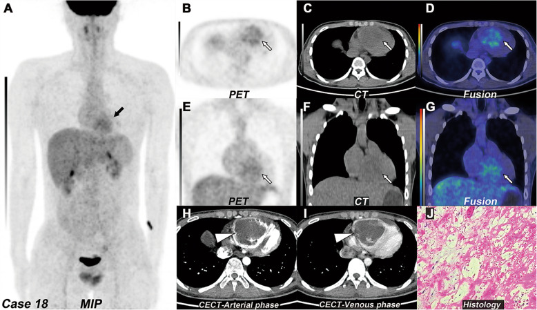
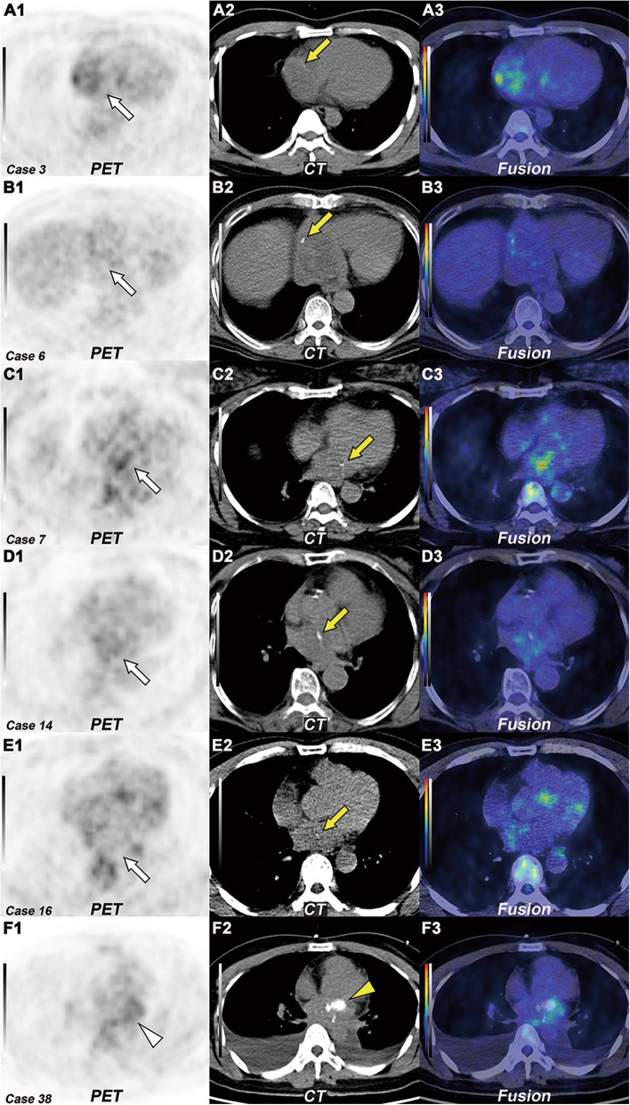
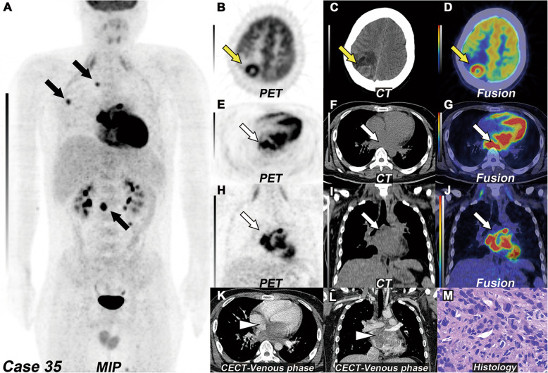
More than half (16/29) of benign tumors exhibited with mild F-FDG uptake. There were significant differences in F-FDG uptake and the degree of absolute enhancement between benign and malignant tumors (P < 0.001). The combination of two modalities improved the specificity from 79 to 93%, the positive predictive value from 73 to 89%, and the accuracy of diagnosis from 85 to 93%. There were significant differences between PET/CT alone or thoracic CECT alone and combined modalities (P = 0.034 and P = 0.026, respectively). The combination with the optimal SUVmax cutoff value generated 94% sensitivity, 100% specificity, 97% negative predictive values, 100% positive predictive values, and 98% accuracy rates.

CONCLUSIONS

Combining F-FDG PET/C with thoracic CECT significantly improved specificity and accuracy compared to CECT or PET/CT alone in detecting tumors. This combination of diagnostic imaging is effective in differentiating malignant from benign masses.

摘要

背景

F-FDG PET/CT是一种用于无创评估和鉴别心脏良恶性肿瘤的关键分子成像模态。然而,很少有良性心脏肿瘤表现为F-FDG摄取增加,这使得鉴别诊断变得困难。本研究旨在回顾性评估F-FDG PET/CT与胸部增强CT(CECT)联合应用,与单独使用CECT或PET/CT相比,是否有助于评估成年患者的原发性心脏肿瘤。

方法

连续纳入46例被诊断为原发性心脏肿瘤的患者。所有患者在活检或手术前均接受F-FDG PET/CT检查,随后进行胸部CECT检查。进行了视觉定性解读和定量分析,并评估了诊断性能。

结果

超过一半(16/29)的良性肿瘤表现为轻度F-FDG摄取。良性和恶性肿瘤在F-FDG摄取和绝对强化程度上存在显著差异(P<0.001)。两种模态联合应用使特异性从79%提高到93%,阳性预测值从73%提高到89%,诊断准确性从85%提高到93%。单独使用PET/CT或胸部CECT与联合模态之间存在显著差异(分别为P = 0.034和P = 0.026)。结合最佳SUVmax临界值可产生94%的灵敏度、100%的特异性、97%的阴性预测值、100%的阳性预测值和98%的准确率。

结论

与单独使用CECT或PET/CT相比,F-FDG PET/C与胸部CECT联合应用在检测肿瘤时显著提高了特异性和准确性。这种诊断成像联合应用在区分恶性和良性肿块方面是有效的。

https://cdn.ncbi.nlm.nih.gov/pmc/blobs/016f/7338301/a31c644bc407/13550_2020_661_Fig7_HTML.jpg
https://cdn.ncbi.nlm.nih.gov/pmc/blobs/016f/7338301/6e94dac4fa3e/13550_2020_661_Fig1_HTML.jpg
https://cdn.ncbi.nlm.nih.gov/pmc/blobs/016f/7338301/929ece5ce9ad/13550_2020_661_Fig2_HTML.jpg
https://cdn.ncbi.nlm.nih.gov/pmc/blobs/016f/7338301/e8dc26b40760/13550_2020_661_Fig3_HTML.jpg
https://cdn.ncbi.nlm.nih.gov/pmc/blobs/016f/7338301/1aee8840a52c/13550_2020_661_Fig4_HTML.jpg
https://cdn.ncbi.nlm.nih.gov/pmc/blobs/016f/7338301/ccb9d3c5fabb/13550_2020_661_Fig5_HTML.jpg
https://cdn.ncbi.nlm.nih.gov/pmc/blobs/016f/7338301/737e31feab2c/13550_2020_661_Fig6_HTML.jpg
https://cdn.ncbi.nlm.nih.gov/pmc/blobs/016f/7338301/a31c644bc407/13550_2020_661_Fig7_HTML.jpg

相似文献

1
Combined PET/CT with thoracic contrast-enhanced CT in assessment of primary cardiac tumors in adult patients.PET/CT联合胸部增强CT在评估成年患者原发性心脏肿瘤中的应用
EJNMMI Res. 2020 Jul 6;10(1):75. doi: 10.1186/s13550-020-00661-x.
2
Diagnosis and staging of cardiac masses: additional value of CMR with F-FDG-PET compared to CMR with CECT.心脏肿块的诊断和分期:与 CECT 相比,CMR 联合 F-FDG-PET 的附加价值。
Eur J Nucl Med Mol Imaging. 2022 Jun;49(7):2232-2241. doi: 10.1007/s00259-022-05709-9. Epub 2022 Mar 5.
3
More advantages in detecting bone and soft tissue metastases from prostate cancer using F-PSMA PET/CT.使用F-PSMA PET/CT检测前列腺癌骨和软组织转移方面有更多优势。
Hell J Nucl Med. 2019 Jan-Apr;22(1):6-9. doi: 10.1967/s002449910952. Epub 2019 Mar 7.
4
Diagnostic and prognostic value of 18F-FDG PET/CT in comparison with morphological imaging in primary adrenal gland malignancies - a multicenter experience.18F-FDG PET/CT与形态学成像相比在原发性肾上腺恶性肿瘤中的诊断和预后价值——一项多中心经验
Hell J Nucl Med. 2015 May-Aug;18(2):97-102. doi: 10.1967/s002449910202. Epub 2015 Jun 29.
5
Evaluation of soft-tissue lesions with (18)F-FDG PET/CT: initial results of a prospective trial.(18)F-FDG PET/CT对软组织病变的评估:一项前瞻性试验的初步结果
Nucl Med Commun. 2014 Mar;35(3):252-9. doi: 10.1097/MNM.0000000000000041.
6
Evaluation of FDG uptake in pulmonary hila with FDG PET/CT and contrast-enhanced CT in patients with thoracic and non-thoracic tumors.评估胸内和非胸内肿瘤患者的 FDG PET/CT 和增强 CT 检查肺门的 FDG 摄取情况。
Ann Nucl Med. 2010 Oct;24(8):593-9. doi: 10.1007/s12149-010-0402-x. Epub 2010 Jul 28.
7
Diagnostic accuracy of (18)F-FDG PET/CT compared with that of contrast-enhanced MRI of the breast at 3 T.3T下(18)F-FDG PET/CT与乳腺对比增强MRI的诊断准确性比较。
Eur J Nucl Med Mol Imaging. 2015 Oct;42(11):1656-1665. doi: 10.1007/s00259-015-3099-1. Epub 2015 Jun 30.
8
FLT PET/CT Is Better Than FDG PET/CT in Differentiating Benign From Malignant Pancreatobiliary Lesions.在鉴别胰腺胆管良性与恶性病变方面,FLT PET/CT比FDG PET/CT更具优势。
Clin Nucl Med. 2016 May;41(5):e244-50. doi: 10.1097/RLU.0000000000001163.
9
Differentiation of malignant and benign cardiac tumors using 18F-FDG PET/CT.应用 18F-FDG PET/CT 鉴别心脏良恶性肿瘤。
J Nucl Med. 2012 Jun;53(6):856-63. doi: 10.2967/jnumed.111.095364. Epub 2012 May 10.
10
Differentiation and diagnosis of benign and malignant testicular lesions using 18F-FDG PET/CT.使用18F-FDG PET/CT对睾丸良恶性病变进行鉴别与诊断。
Eur J Radiol. 2017 Aug;93:114-120. doi: 10.1016/j.ejrad.2017.05.001. Epub 2017 May 26.

引用本文的文献

1
[F]FDG PET imaging in the differentiation of cardiac masses: an updated systematic review and dual Meta-Analysis of diagnostic performance and parameter variability.[F]氟代脱氧葡萄糖正电子发射断层扫描成像在心脏肿块鉴别诊断中的应用:诊断性能及参数变异性的最新系统评价与双元荟萃分析
Eur J Nucl Med Mol Imaging. 2025 Apr 29. doi: 10.1007/s00259-025-07289-w.
2
COVID-19 associated mucormycosis: evolving technologies for early and rapid diagnosis.新型冠状病毒肺炎相关毛霉病:早期快速诊断的技术进展
3 Biotech. 2022 Jan;12(1):6. doi: 10.1007/s13205-021-03080-4. Epub 2021 Dec 6.
3
Diagnostic challenges in primary cardiac lymphoma, the opportunity of F-FDG PET/CT integrated with contrast-enhanced CT.

本文引用的文献

1
Impact of optimized PET imaging conditions on F-FDG uptake quantification in patients with apparently normal aortas.优化 PET 成像条件对主动脉外观正常患者 F-FDG 摄取定量的影响。
J Nucl Cardiol. 2021 Aug;28(4):1349-1359. doi: 10.1007/s12350-019-01833-6. Epub 2019 Aug 6.
2
Primary cardiac fibroma with persistent left superior vena cava in a young adult: Contrast-enhanced CT and F-FDG PET/CT finding.一名年轻成人原发性心脏纤维瘤合并永存左上腔静脉:对比增强CT及F-FDG PET/CT表现
J Nucl Cardiol. 2020 Oct;27(5):1837-1840. doi: 10.1007/s12350-019-01825-6. Epub 2019 Jul 25.
3
Total-Body Dynamic Reconstruction and Parametric Imaging on the uEXPLORER.
原发性心脏淋巴瘤的诊断挑战:F-FDG PET/CT 与增强 CT 结合的机遇。
J Nucl Cardiol. 2022 Oct;29(5):2378-2389. doi: 10.1007/s12350-021-02723-6. Epub 2021 Jul 27.
uEXPLORER 全身动态重建与参数成像。
J Nucl Med. 2020 Feb;61(2):285-291. doi: 10.2967/jnumed.119.230565. Epub 2019 Jul 13.
4
Recommendations for F-fluorodeoxyglucose positron emission tomography imaging for diagnosis of cardiac sarcoidosis-2018 update: Japanese Society of Nuclear Cardiology recommendations.2018年更新版:日本核心脏病学会关于¹⁸F-氟脱氧葡萄糖正电子发射断层显像用于心脏结节病诊断的建议
J Nucl Cardiol. 2019 Aug;26(4):1414-1433. doi: 10.1007/s12350-019-01755-3.
5
F-FDG-PET/CT imaging in cardiac tumors: illustrative clinical cases and review of the literature.心脏肿瘤的F-FDG-PET/CT成像:典型临床病例及文献综述
Ther Adv Med Oncol. 2018 Aug 30;10:1758835918793569. doi: 10.1177/1758835918793569. eCollection 2018.
6
Right atrial epithelioid angiosarcoma with multiple pulmonary metastasis confirmed by multimodality imaging-guided pulmonary biopsy: A case report and literature review.多模态成像引导下肺活检确诊的右房上皮样血管肉瘤伴多发肺转移:病例报告及文献复习
Medicine (Baltimore). 2018 Jul;97(30):e11588. doi: 10.1097/MD.0000000000011588.
7
Diagnostic accuracy and prognostic value of simultaneous hybrid 18F-fluorodeoxyglucose positron emission tomography/magnetic resonance imaging in cardiac sarcoidosis.18F-氟脱氧葡萄糖正电子发射断层扫描/磁共振成像在心脏结节病中的诊断准确性和预后价值。
Eur Heart J Cardiovasc Imaging. 2018 Jul 1;19(7):757-767. doi: 10.1093/ehjci/jex340.
8
What is the role of 18F-FDG uptake intensity in suspected atrial myxoma exploration?18F-FDG摄取强度在疑似心房黏液瘤探查中的作用是什么?
J Nucl Cardiol. 2018 Oct;25(5):1861-1862. doi: 10.1007/s12350-017-1094-6. Epub 2017 Oct 30.
9
Radiological characteristics of atrial myxoma in Cardiac Computed Tomography.心脏计算机断层扫描中心房黏液瘤的放射学特征
J Cardiovasc Comput Tomogr. 2017 May-Jun;11(3):234-236. doi: 10.1016/j.jcct.2017.02.002. Epub 2017 Feb 7.
10
PET/CT-guided percutaneous biopsy of FDG-avid metastatic bone lesions in patients with advanced lung cancer: a safe and effective technique.PET/CT引导下对晚期肺癌患者FDG摄取阳性的转移性骨病变进行经皮活检:一种安全有效的技术。
Eur J Nucl Med Mol Imaging. 2017 Jan;44(1):25-32. doi: 10.1007/s00259-016-3455-9. Epub 2016 Jul 22.