Department of Oncology, University of Alberta, Edmonton, AB, Canada.
Department of Pathology and Laboratory Medicine, Foothills Medical Centre, University of Calgary, Calgary, AB, Canada.
Front Immunol. 2020 Jun 19;11:1287. doi: 10.3389/fimmu.2020.01287. eCollection 2020.
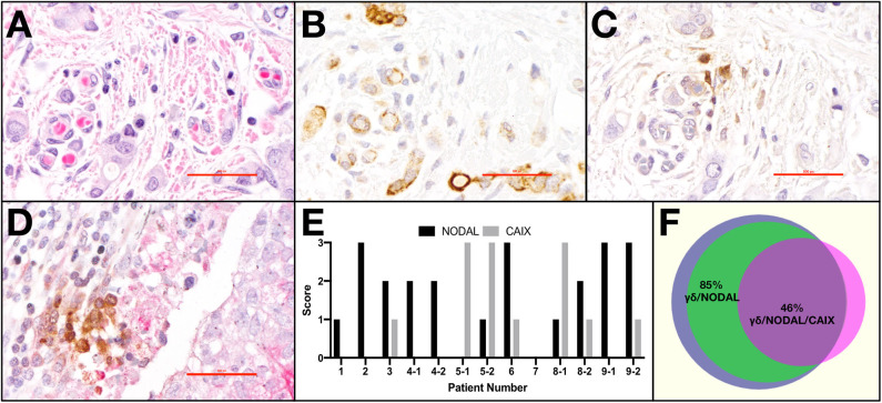
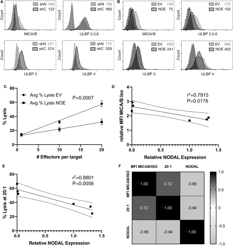
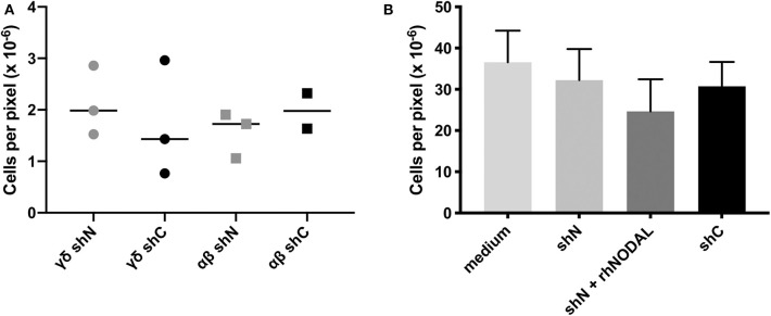
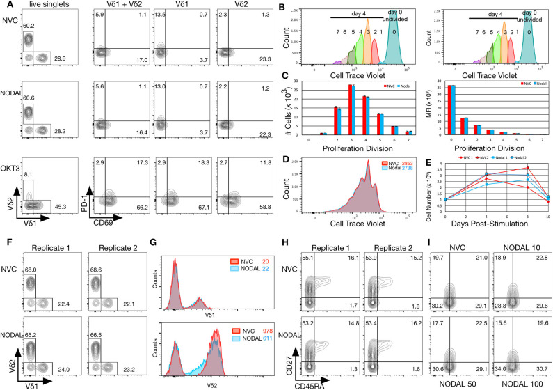
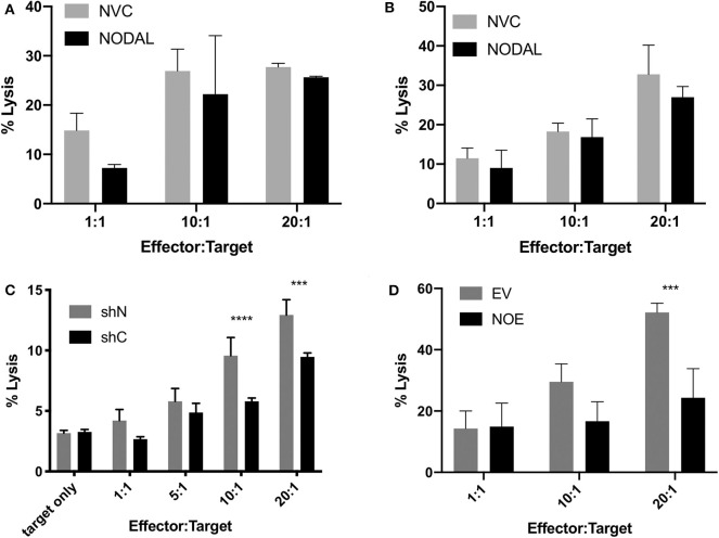
Gamma delta (γδ) T cells kill transformed cells, and increased circulating γδ T cells levels correlate with improved outcome in cancer patients; however, their function within the breast tumor microenvironment (TME) remains controversial. As tumors progress, they begin to express stem-cell associated proteins, concomitant with the emergence of therapy resistant metastatic disease. For example, invasive breast cancers often secrete the embryonic morphogen, NODAL. NODAL has been shown to promote angiogenesis, therapy resistance and metastasis in breast cancers. However, to date, little is known about how this secreted protein may interact with cells in the TME. Herein we explore how NODAL in the TME may influence γδ T cell function. We have assessed the proximity of γδ T cells to NODAL in a cohort of triple negative breast tumors. In all cases in which γδ T cells could be identified in these tumors, γδ T cells were found in close proximity to NODAL-expressing tumor cells. Migration of γδ and αβ T cells was similar toward MDA-MB-231 cells in which NODAL had been knocked down (shN) and MDA-MB-231 scrambled control cells (shC). Furthermore, Vδ1 γδ T cells did not migrate preferentially toward conditioned medium from these cell lines. While 24-h exposure to NODAL did not impact CD69, PD-1, or T cell antigen receptor (TCR) expression on γδ T cells, long term exposure resulted in decreased Vδ2 TCR expression. Maturation of γδ T cells was not significantly influenced by NODAL stimulation. While neither short- nor long-term NODAL stimulation impacted the ability of γδ T cells to kill MCF-7 breast cancer cells, the absence of NODAL resulted in greater sensitivity of targets to γδ T cell cytotoxicity, while overexpression of NODAL conferred resistance. This appeared to be at least in part due to an inverse correlation between NODAL and surface MICA/B expression on breast cancer target lines. As such, it appears that NODAL may play a role in strategies employed by breast cancer cells to evade γδ T cell targeting, and this should be considered in the development of safe and effective γδ T cell immunotherapies.
γδ(γδ)T 细胞可以杀死转化细胞,并且循环 γδ T 细胞水平的增加与癌症患者的预后改善相关;然而,它们在乳腺肿瘤微环境(TME)中的功能仍存在争议。随着肿瘤的进展,它们开始表达与干细胞相关的蛋白质,同时出现治疗耐药性转移性疾病。例如,侵袭性乳腺癌通常分泌胚胎形态发生素 NODAL。已经表明,NODAL 可促进乳腺癌的血管生成、治疗耐药性和转移。然而,迄今为止,人们对这种分泌蛋白如何与 TME 中的细胞相互作用知之甚少。在此,我们探讨了 TME 中的 NODAL 如何影响 γδ T 细胞的功能。我们评估了三阴性乳腺癌患者队列中 NODAL 与 γδ T 细胞的接近程度。在这些肿瘤中可以识别 γδ T 细胞的所有情况下,都发现 γδ T 细胞与表达 NODAL 的肿瘤细胞密切相邻。NODAL 敲低(shN)和 MDA-MB-231 对照细胞(shC)中 MDA-MB-231 细胞中 γδ 和 αβ T 细胞的迁移相似。此外,Vδ1 γδ T 细胞不会优先向这些细胞系的条件培养基中迁移。虽然 NODAL 暴露 24 小时不会影响 γδ T 细胞上的 CD69、PD-1 或 T 细胞抗原受体(TCR)表达,但长期暴露会导致 Vδ2 TCR 表达减少。NODAL 刺激对 γδ T 细胞的成熟没有显著影响。虽然 NODAL 的短期或长期刺激都不会影响 γδ T 细胞杀伤 MCF-7 乳腺癌细胞的能力,但 NODAL 的缺失会导致靶细胞对 γδ T 细胞细胞毒性的敏感性增加,而过表达 NODAL 则会导致耐药性。这似乎至少部分是由于乳腺癌靶细胞系中 NODAL 与表面 MICA/B 表达之间的反比关系。因此,NODAL 似乎在乳腺癌细胞逃避 γδ T 细胞靶向的策略中发挥作用,在开发安全有效的 γδ T 细胞免疫疗法时应考虑这一点。