Melendez Patricia E, Nguyen Trinh T, Bhatt Alok A, Kaproth-Joslin Katherine
University of Rochester Medical Center, 601 Elmwood Ave, Box 648, Rochester, NY, 14642, USA.
Mayo Clinic, 4500 San Pablo Road, Jacksonville, FL, 32224, USA.
Insights Imaging. 2020 Jul 8;11(1):82. doi: 10.1186/s13244-020-00879-2.
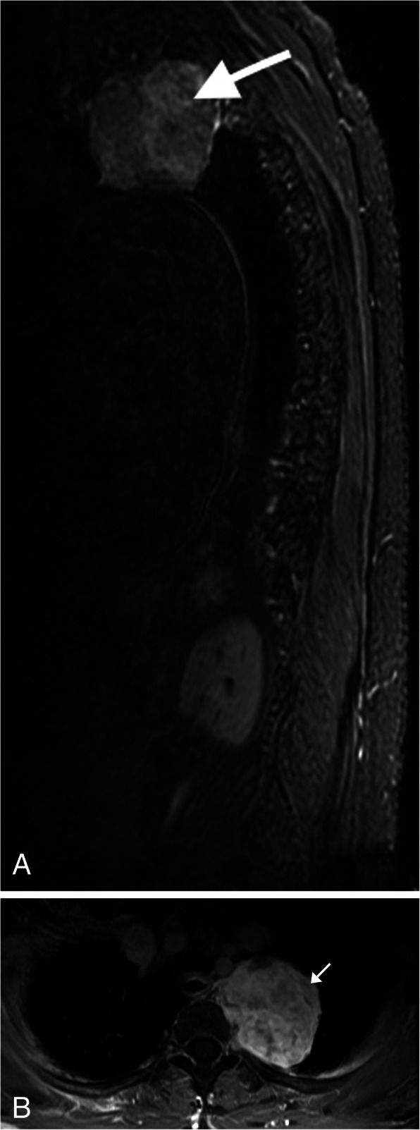
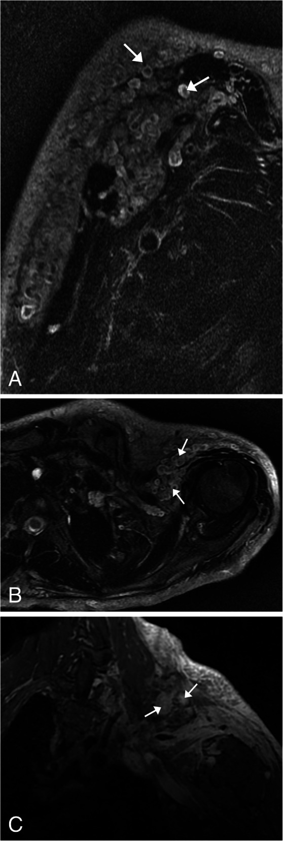
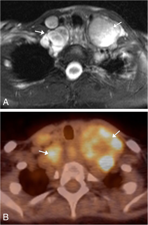
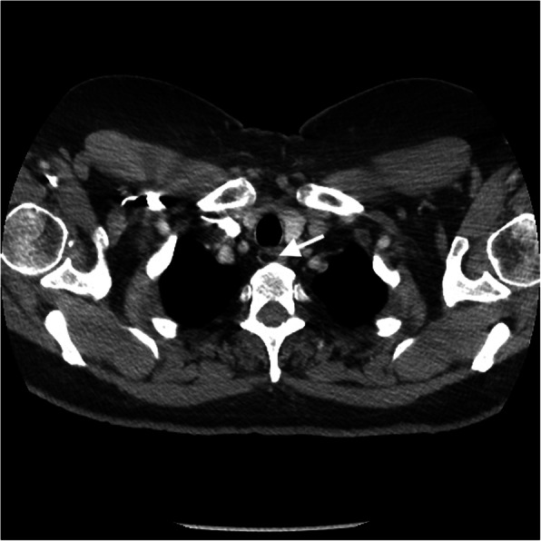
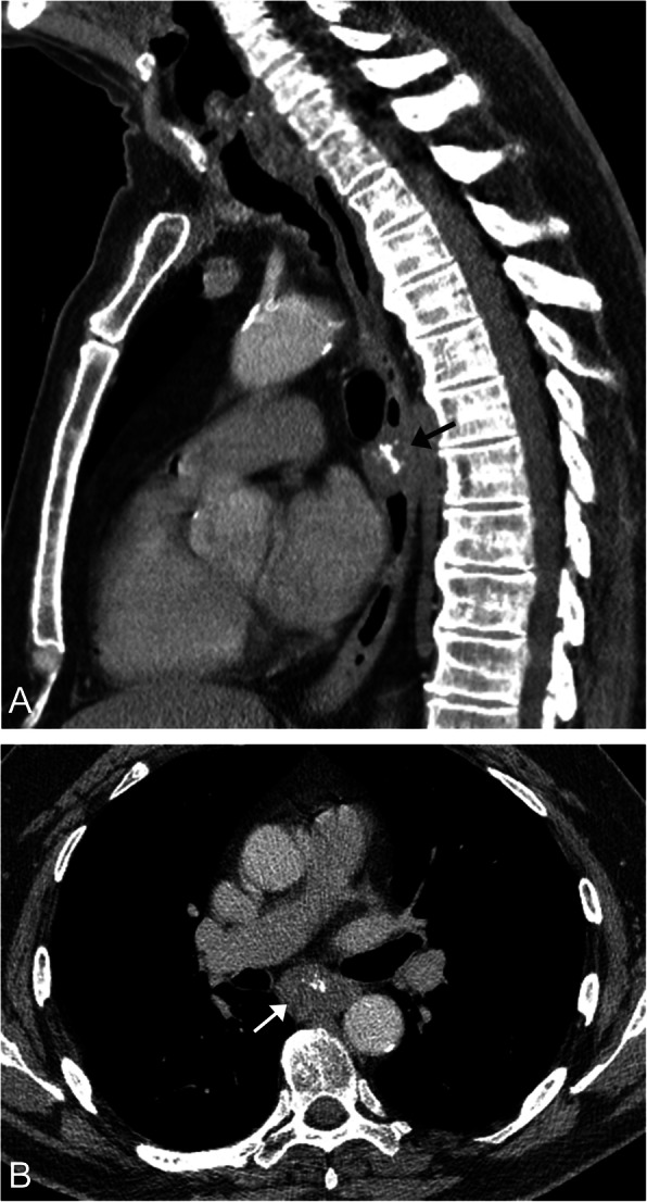
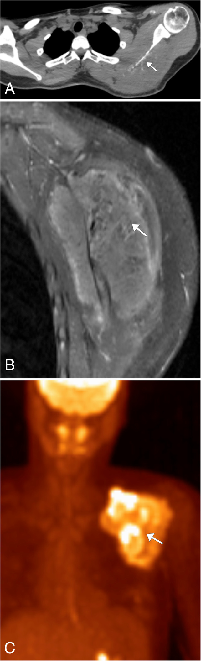
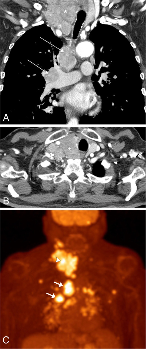
The thoracic inlet is located at the crossroads between imaging of the neck and the chest. Its location is an important anatomic landmark, serving as the central conducting pathway for many vital structures extending from the neck into the chest and vice versa. Many critical body systems, including the respiratory, lymphatic, neurologic, enteric, musculoskeletal, endocrine, and vascular systems, are located within this region. Neoplasms, both benign and malignant, can arise in any of the body systems located in this area. Due to the small size of this anatomic location, pathology is easily overlooked and imagers should be aware of the imaging appearance of these neoplasms, as well as which imaging modality is the most appropriate for neoplasm evaluation. This article will present an image rich, system-based discussion of the neoplastic pathology that can occur in this region. The anatomy of the thoracic inlet and the non-neoplastic pathology of the thoracic inlet have been covered in our companion article.
胸廓入口位于颈部和胸部成像的交叉点。其位置是一个重要的解剖标志,是许多从颈部延伸至胸部及反之亦然的重要结构的中央传导通路。包括呼吸、淋巴、神经、肠道、肌肉骨骼、内分泌和血管系统在内的许多关键身体系统都位于该区域。良性和恶性肿瘤都可能出现在该区域的任何身体系统中。由于这个解剖位置较小,病变很容易被忽视,影像科医生应了解这些肿瘤的影像表现,以及哪种成像方式最适合肿瘤评估。本文将基于系统,对该区域可能发生的肿瘤性病变进行图文并茂的讨论。我们的配套文章已涵盖胸廓入口的解剖结构和胸廓入口的非肿瘤性病变。