Department of Pediatric Cardiology and Cardiovascular Surgery, Bambino Gesù Children's Research Hospital-IRCSS, Piazza Sant'Onofrio 4, 00165, Rome, Italy.
Department of Imaging, Bambino Gesù Children's Research Hospital-IRCSS, Rome, Italy.
Sci Rep. 2020 Jul 9;10(1):11321. doi: 10.1038/s41598-020-68048-5.
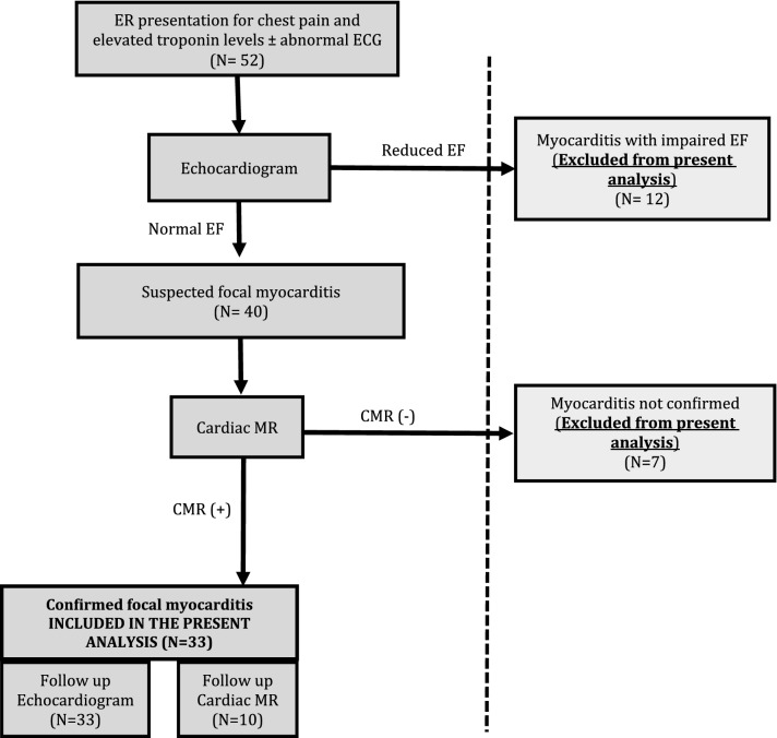
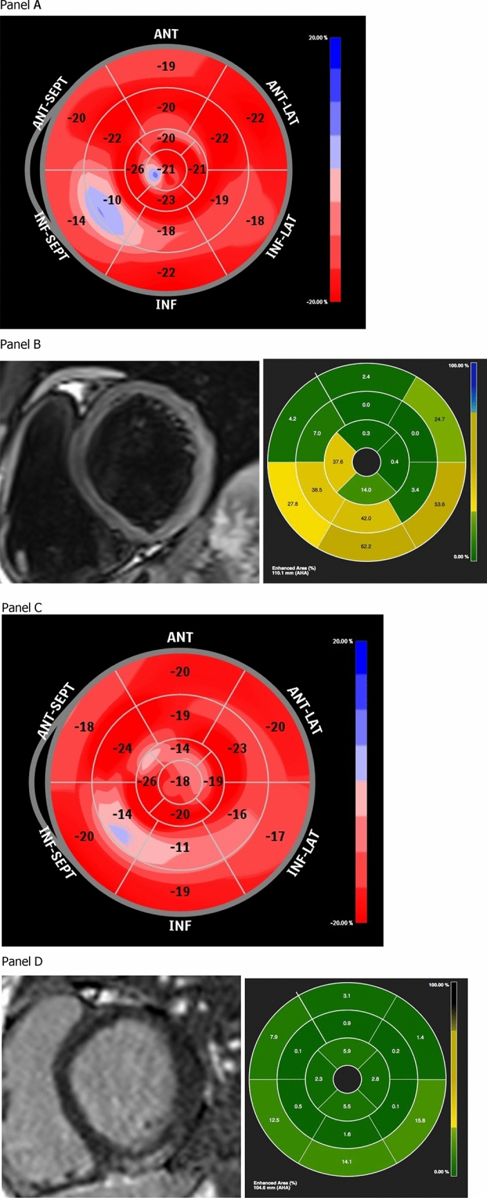
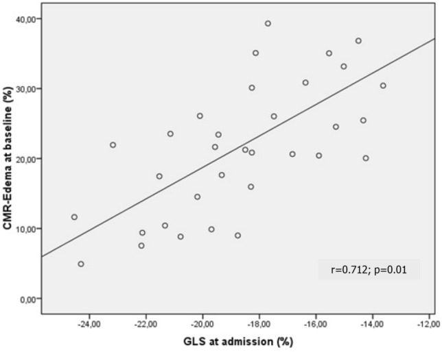
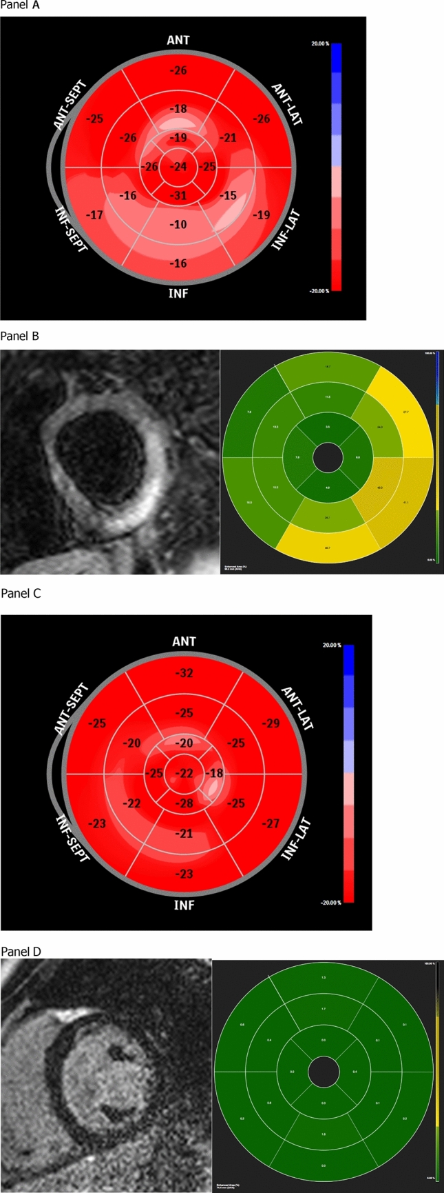
The aim here was to describe the role of speckle tracking echocardiography (STE), in identifying impairment in systolic function in children and adolescents with focal myocarditis and without reduction in ejection fraction. We describe data from 33 pediatric patients (age 4-17 years) admitted for focal myocarditis, confirmed by cardiac magnetic resonance (CMR), and without impaired ejection fraction and/or wall motion abnormalities. All children underwent Doppler echocardiography examination with analysis of global (G) and segmental longitudinal strain (LS) and CMR for the quantification of edema and myocardial fibrosis. Reduction in LS was defined according to age-specific partition values. At baseline, impaired GLS was present in 58% of patients (n = 19), albeit normal ejection fraction. LS was also regionally impaired, according to the area of higher edema at CMR (i.e. most impaired at the level of the infero-lateral segments as compared to other segments (p < 0.05). GLS impairment was also moderately correlated with the percentage edema at CMR (r = - 0.712; p = 0.01). At follow-up, GLS improved in all patients (p < 0.001), and normal values were found in 13/19 patients with baseline reduction. Accordingly persistent global and regional impairment was still observed in 6 patients. Patients with persistent LS reduction demonstrated residual focal cardiac fibrosis at follow-up CMR. Both global and regional LS is able to identify abnormalities in systolic longitudinal mechanics in children and adolescents with focal myocarditis and normal ejection fraction. The reduction in LS is consistent with edema amount and localization at CMR. Furthermore, LS identifies regional recovery or persistent cardiac function impairment, possibly related to residual focal fibrosis.
本研究旨在描述斑点追踪超声心动图(STE)在识别局灶性心肌炎且射血分数无降低的儿童和青少年收缩功能障碍中的作用。我们描述了 33 名儿科患者(年龄 4-17 岁)的数据,这些患者因局灶性心肌炎住院,经心脏磁共振(CMR)证实,且射血分数和/或壁运动异常无受损。所有儿童均接受了多普勒超声心动图检查,分析了整体(G)和节段性纵向应变(LS),并进行了 CMR 检查以量化水肿和心肌纤维化。根据年龄特异性分区值,LS 降低定义为 LS 降低。在基线时,尽管射血分数正常,但仍有 58%的患者(n=19)存在 GLS 受损。LS 也存在区域性受损,根据 CMR 中水肿面积较大的区域(即下外侧节段较其他节段受损更严重(p<0.05)。GLS 受损与 CMR 中水肿百分比也呈中度相关(r=-0.712;p=0.01)。在随访时,所有患者的 GLS 均有所改善(p<0.001),且在基线时降低的 19 名患者中有 13 名恢复正常。因此,仍有 6 名患者持续存在 GLS 受损。在随访 CMR 时,持续存在 LS 降低的患者存在残余局灶性心脏纤维化。GLS 和区域性 LS 均能够识别射血分数正常的局灶性心肌炎儿童和青少年的收缩纵向力学异常。LS 降低与 CMR 中的水肿量和定位一致。此外,LS 可识别区域恢复或持续的心脏功能障碍,可能与残余局灶性纤维化有关。