Department of Orthopaedic Surgery, Surgical Science, Tokai University School of Medicine, Isehara, Japan.
Center for Musculoskeletal innovative Research and Advancement (C-MiRA), Tokai University Graduate School, Isehara, Japan.
J Tissue Eng Regen Med. 2020 Sep;14(9):1296-1306. doi: 10.1002/term.3101. Epub 2020 Jul 22.
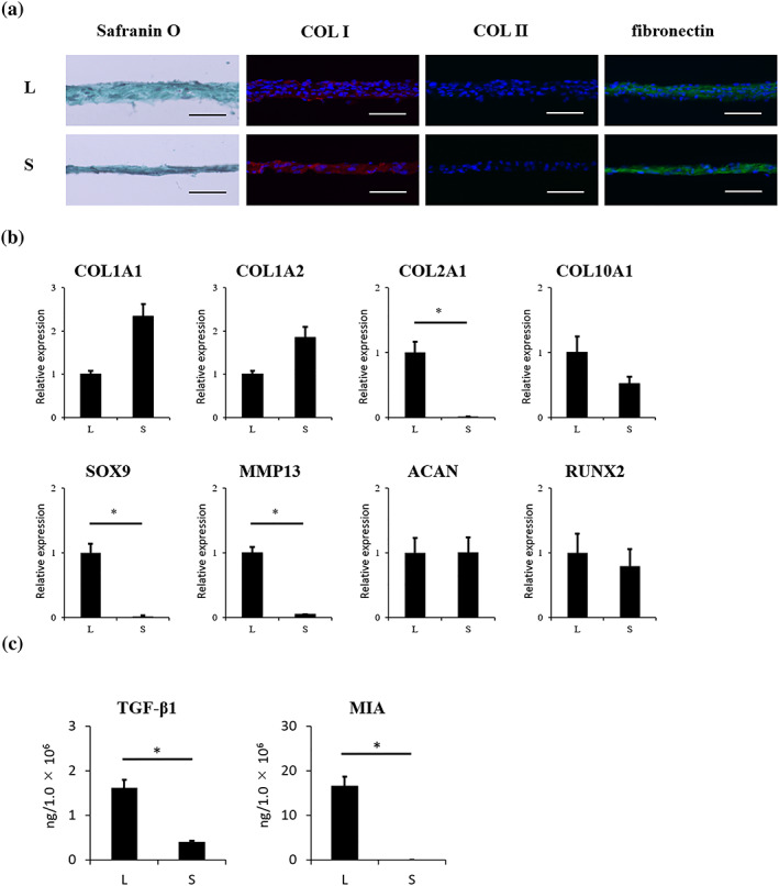
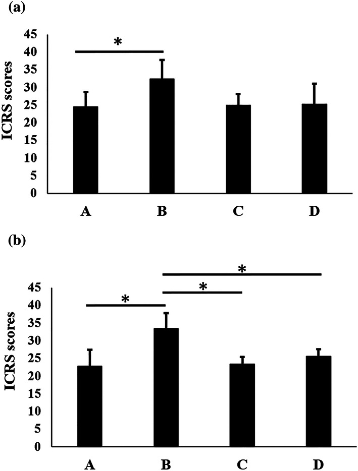
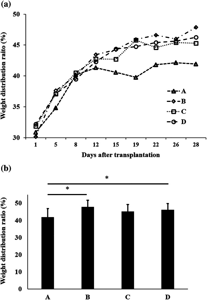
Although cell transplantation has attracted much attention in regenerative medicine, animal models continue to be used in translational research to evaluate safety and efficacy because cell sources and transplantation modalities are so diverse. In the present study, we investigated the regenerative effects of human chondrocyte sheets on articular cartilage in a xenogeneic transplantation model using immune-deficient rats. Osteochondral defects were created in the knee joints of immune-deficient rats that were treated as Group A, untreated (without transplantation); Group B, transplantation of a layered chondrocyte sheet containing 5.0 × 10 cells (layered chondrocyte sheet transplantation); Group C, transplantation of a synoviocyte sheet containing 5.0 × 10 cells (synoviocyte sheet transplantation); or Group D, transplantation of both a synoviocyte sheet plus a layered chondrocyte sheet, each containing 5.0 × 10 cells (synoviocyte sheet plus layered chondrocyte sheet transplantation). Histological evaluation demonstrated that Group B showed cartilage regeneration with hyaline cartilage and fibrocartilage. In Groups C and D, the defect was filled with fibrous tissue but no hyaline cartilage. Transplanted cells were detected at 4 and 12 weeks after transplantation, but the number of cells had decreased at 12 weeks. Our results indicate that layered chondrocyte sheet transplantation contributes to articular cartilage regeneration; this model proved useful for evaluating these regenerative effects.
尽管细胞移植在再生医学中引起了广泛关注,但动物模型仍被用于转化研究,以评估安全性和疗效,因为细胞来源和移植方式多种多样。在本研究中,我们使用免疫缺陷大鼠的异种移植模型研究了人软骨细胞片对关节软骨的再生作用。在免疫缺陷大鼠的膝关节中创建了骨软骨缺损,将其分为 A 组(未处理,未移植)、B 组(移植 5.0×10 个细胞的层状软骨细胞片)、C 组(移植 5.0×10 个细胞的滑膜细胞片)或 D 组(分别移植 5.0×10 个细胞的滑膜细胞片和层状软骨细胞片)。组织学评估表明,B 组表现出透明软骨和纤维软骨的软骨再生。在 C 组和 D 组中,缺陷部位填充了纤维组织,但没有透明软骨。在移植后 4 周和 12 周检测到移植细胞,但在 12 周时细胞数量减少。我们的结果表明,层状软骨细胞片移植有助于关节软骨再生;该模型对于评估这些再生效果非常有用。