Liu Ling-Ling, Qiao Shan, Wang Mei-Ling, Wu Huai-Kuan, Su Yong-Xin, Wang Ke-Mo, Liu Xue-Wu
Department of Neurology, Qilu Hospital, Cheeloo College of Medicine, Shandong University, Jinan, China.
Department of Neurology, Liaocheng People's Hospital, Liaocheng, China.
Front Neurosci. 2020 Jun 25;14:613. doi: 10.3389/fnins.2020.00613. eCollection 2020.
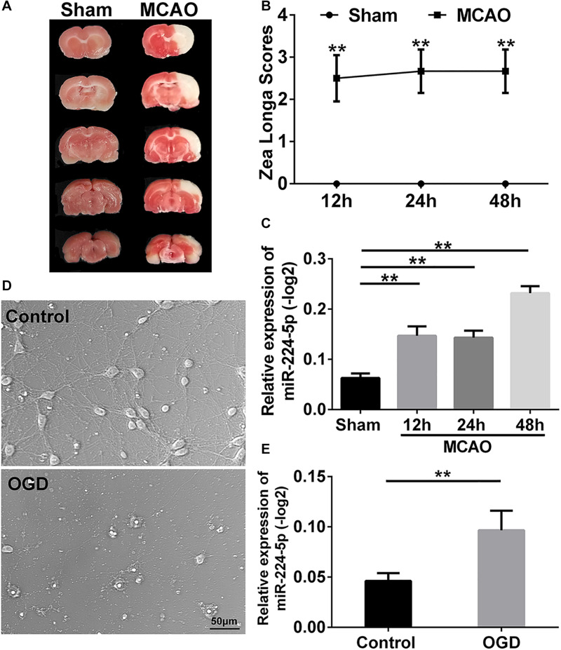
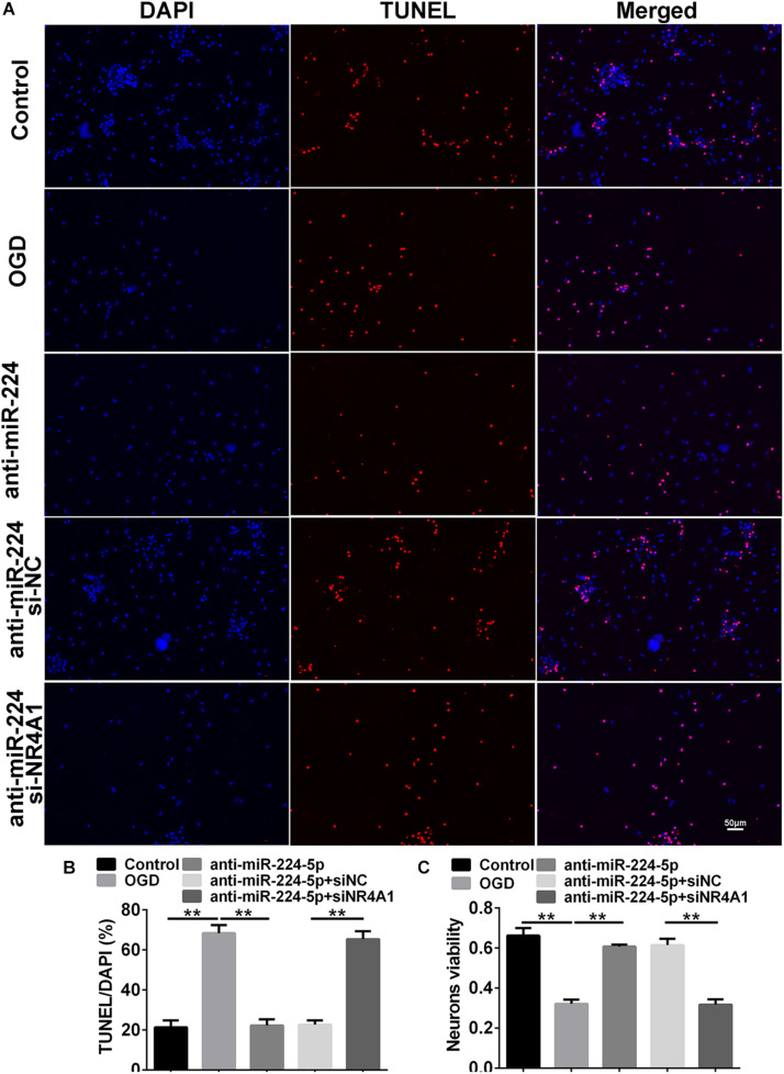
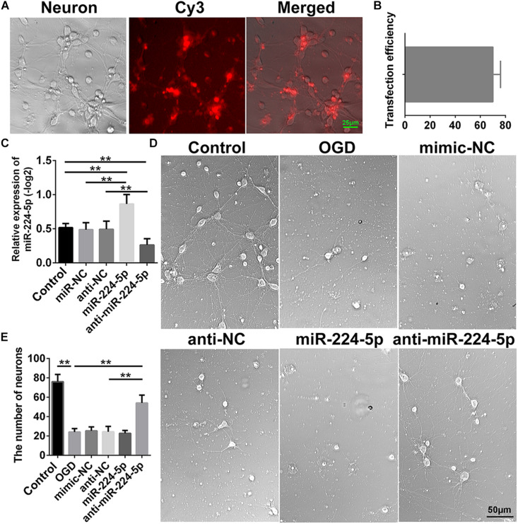
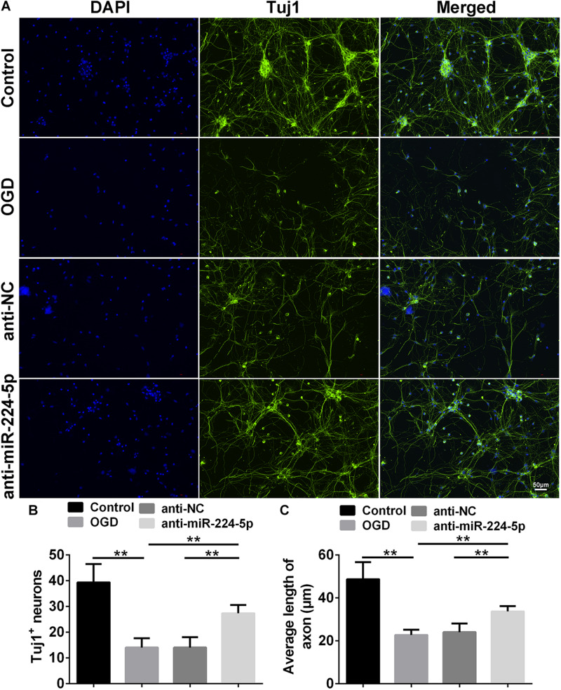
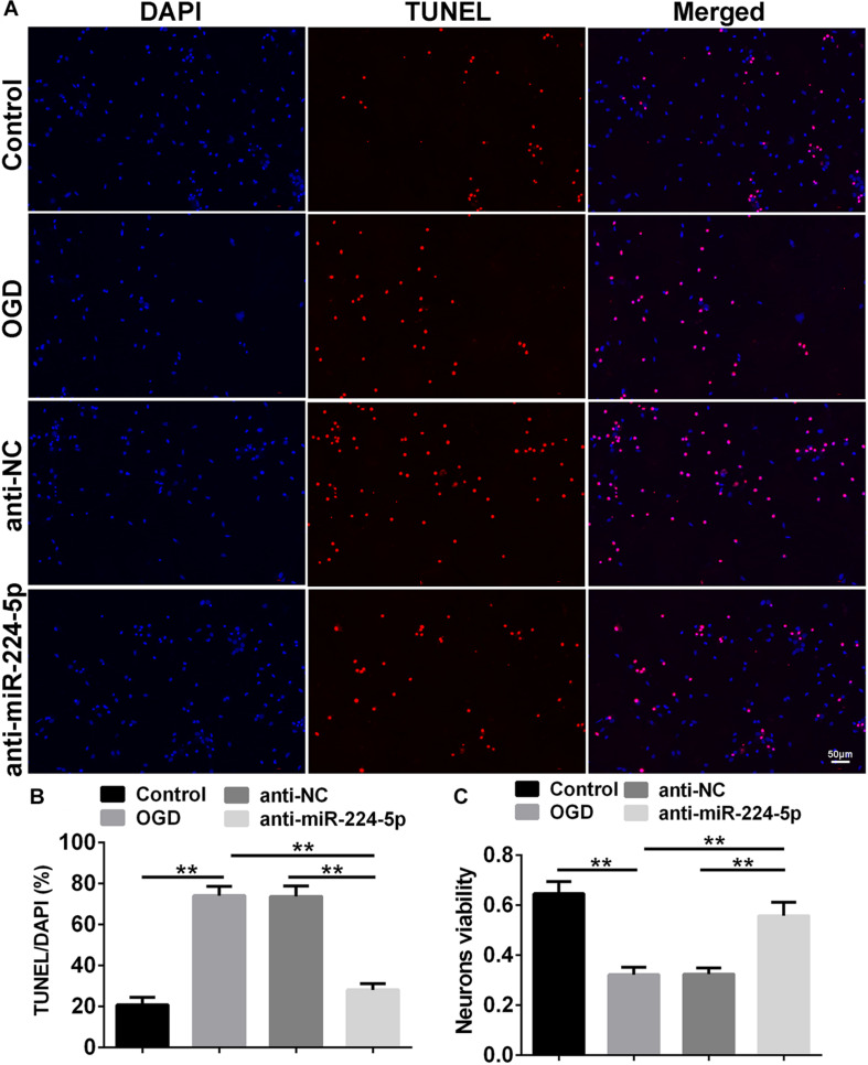
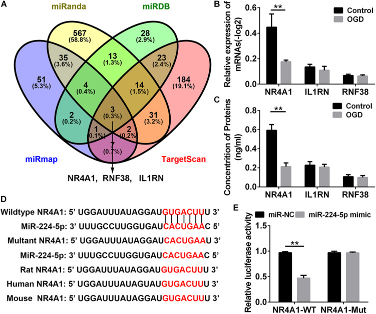
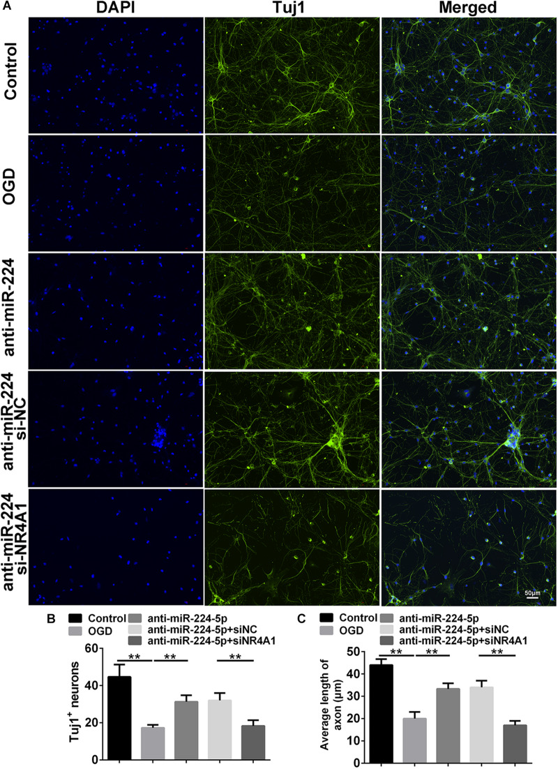
This study was designed to investigate the molecular mechanism of stroke and to explore the effect of miR-224-5p in hypoxic cortical neurons. Firstly, we established a middle cerebral artery occlusion (MCAO) model with Sprague-Dawley rats. Triphenyltetrazolium chloride (TTC) staining showed the brain infarction of an MCAO rat. Longa scores of rats were significantly increased in 12th, 24th, and 48th hours after MCAO. Then, we found that miR-224-5p was increased after MCAO in rats by qRT-PCR. In order to investigate the effect of miR-224-5p in hypoxic neurons, we established an oxygen-glucose deprivation (OGD) model with cortical neurons. MiR-224-5p was also upregulated in neurons after OGD by qRT-PCR. After transfection of the miR-224-5p inhibitor, the number of neurons in the anti-miR-224-5p group significantly increased ( < 0.01) in comparison to the anti-NC group. Furthermore, Tuj1 (neuronal marker) staining and TUNEL assay (to detect apoptotic cells) were performed in neurons. The survival of neurons in the anti-miR-224-5p group was significantly improved ( < 0.01), while the apoptosis of neurons in the anti-miR-224-5p group was significantly decreased ( < 0.01), when compared with that of the anti-NC group. In addition, we predicted that potential target genes of miR-224-5p were nuclear receptor subfamily 4 group A member 1 (NR4A1), interleukin 1 receptor antagonist (IL1RN), and ring finger protein 38 (RNF38) with bioinformatics databases, such as TargetScan, miRDB, miRmap, and miRanda. The result of qRT-PCR confirmed that NR4A1 was significantly decreased after hypoxic injury ( < 0.01). Meanwhile, luciferase reporter's assay indicated that NR4A1 was the direct target of miR-224-5p. Compared with the anti-miR-224-5p + siNC group, the number of cortical neurons and the length of the neuron axon in the anti-miR-224-5p + si-NR4A1 group were significantly decreased ( < 0.01), and the number of neuronal apoptosis in the anti-miR-224-5p + si-NR4A1 group was increased ( < 0.01). In conclusion, miR-224-5p played a crucial role in hypoxic neuron injury through NR4A1, which might be an important regulatory mechanism in OGD injury of neurons.
本研究旨在探讨中风的分子机制,并探究miR-224-5p在缺氧皮质神经元中的作用。首先,我们用Sprague-Dawley大鼠建立了大脑中动脉闭塞(MCAO)模型。氯化三苯基四氮唑(TTC)染色显示了MCAO大鼠的脑梗死情况。MCAO术后12小时、24小时和48小时,大鼠的Longa评分显著升高。然后,我们通过qRT-PCR发现MCAO后大鼠体内miR-224-5p水平升高。为了研究miR-224-5p在缺氧神经元中的作用,我们用皮质神经元建立了氧糖剥夺(OGD)模型。通过qRT-PCR也发现OGD后神经元中miR-224-5p上调。转染miR-224-5p抑制剂后,与抗NC组相比,抗miR-224-5p组的神经元数量显著增加(<0.01)。此外,对神经元进行了Tuj1(神经元标志物)染色和TUNEL检测(用于检测凋亡细胞)。与抗NC组相比,抗miR-224-5p组神经元的存活率显著提高(<0.01),而抗miR-224-5p组神经元的凋亡率显著降低(<0.01)。另外,我们利用TargetScan、miRDB、miRmap和miRanda等生物信息学数据库预测miR-224-5p的潜在靶基因是核受体亚家族4A组成员1(NR4A1)、白细胞介素1受体拮抗剂(IL1RN)和环指蛋白38(RNF38)。qRT-PCR结果证实缺氧损伤后NR4A1显著降低(<0.01)。同时,荧光素酶报告基因检测表明NR4A1是miR-224-5p的直接靶标。与抗miR-224-5p+siNC组相比,抗miR-224-5p+si-NR4A1组皮质神经元的数量和神经元轴突的长度显著减少(<0.01),抗miR-224-5p+si-NR4A1组神经元凋亡数量增加(<0.01)。总之,miR-224-5p通过NR4A1在缺氧神经元损伤中起关键作用,这可能是神经元OGD损伤的重要调控机制。