Department of Psychology, Florida State University, Tallahassee, FL 32304
Department of Psychology, Florida State University, Tallahassee, FL 32304.
eNeuro. 2020 Jul 31;7(4). doi: 10.1523/ENEURO.0053-20.2020. Print 2020 Jul/Aug.
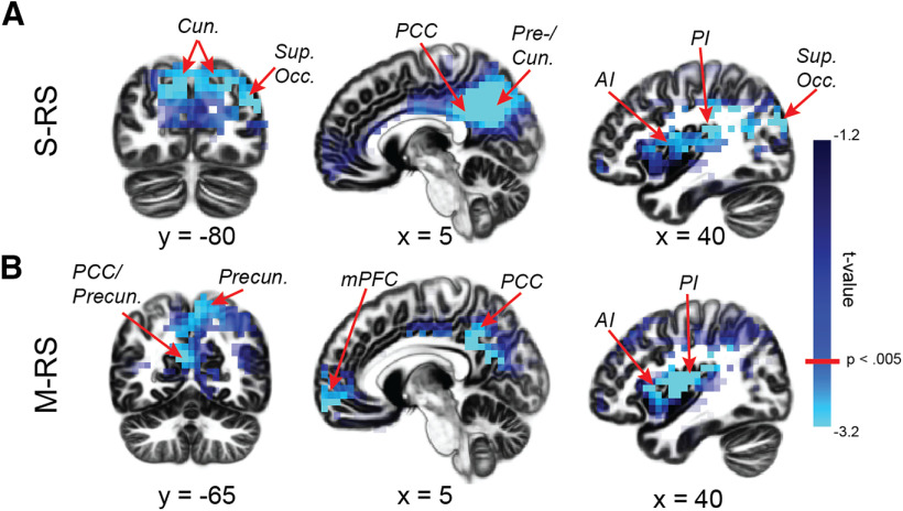
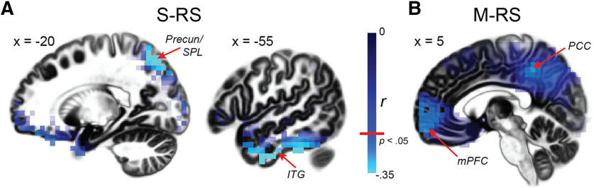
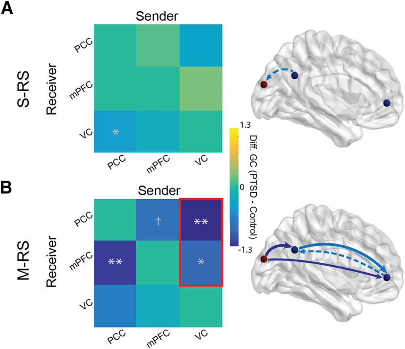
Anomalies in default mode network (DMN) activity and α (8-12 Hz) oscillations have been independently observed in posttraumatic stress disorder (PTSD). Recent spatiotemporal analyses suggest that α oscillations support DMN functioning via interregional synchronization and sensory cortical inhibition. Therefore, we examined a unifying pathology of α deficits in the visual-cortex-DMN system in PTSD. Human patients with PTSD (=25) and two control groups, patients with generalized anxiety disorder (GAD; =24) and healthy controls (HCs; =20), underwent a standard eyes-open resting state (S-RS) and a modified resting state (M-RS) of passively viewing salient images (known to deactivate the DMN). High-density electroencephalogram (hdEEG) were recorded, from which intracortical α activity (power and connectivity/Granger causality) was extracted using the exact low-resolution electromagnetic tomography (eLORETA). Patients with PTSD (vs GAD/HC) demonstrated attenuated α power in the visual cortex (VC) and key hubs of the DMN [posterior cingulate cortex (PCC) and medial prefrontal cortex (mPFC)] at both states, the severity of which further correlated with hypervigilance symptoms. With increased visual input (at M-RS vs S-RS), patients with PTSD further demonstrated reduced α-frequency directed connectivity within the DMN (PCC→mPFC) and, importantly, from the VC to both DMN hubs (VC→PCC and VC→mPFC), linking α deficits in the two systems. These interrelated α deficits align with DMN hypoactivity/hypoconnectivity, sensory disinhibition, and hypervigilance in PTSD, representing a unifying neural underpinning of these anomalies. The identification of visual-cortex-DMN α dysrhythmia in PTSD further presents a novel therapeutic target, promoting network-based intervention of neural oscillations.
在创伤后应激障碍(PTSD)中,已经独立观察到默认模式网络(DMN)活动和α(8-12Hz)振荡的异常。最近的时空分析表明,α振荡通过区域间同步和感觉皮质抑制来支持 DMN 功能。因此,我们研究了 PTSD 中视觉皮层-DMN 系统中α功能缺陷的统一病理学。人类 PTSD 患者(n=25)和两个对照组,广泛性焦虑障碍(GAD;n=24)和健康对照组(HC;n=20)接受了标准睁眼静息状态(S-RS)和被动观看显著图像的修改静息状态(M-RS)(已知激活 DMN)。记录高密度脑电图(hdEEG),从中使用精确低分辨率电磁断层成像(eLORETA)提取皮质内α活动(功率和连接/格兰杰因果关系)。与 GAD/HC 相比,PTSD 患者在 S-RS 和 M-RS 状态下,视觉皮层(VC)和 DMN 的关键中枢(后扣带回皮层(PCC)和内侧前额叶皮层(mPFC))的α功率减弱,其严重程度与过度警觉症状进一步相关。随着视觉输入的增加(在 M-RS 与 S-RS 相比),PTSD 患者进一步表现出 DMN 内α频率定向连接减少(PCC→mPFC),并且重要的是,从 VC 到两个 DMN 中枢(VC→PCC 和 VC→mPFC),将两个系统中的α缺陷联系起来。这些相互关联的α缺陷与 PTSD 中的 DMN 活动减少/连接减少、感觉抑制和过度警觉一致,代表这些异常的统一神经基础。在 PTSD 中识别出视觉皮层-DMN α节律紊乱进一步提出了一个新的治疗靶点,促进基于网络的神经振荡干预。