Institute for Molecular Bioscience, University of Queensland, Brisbane, QLD, 4072, Australia.
Electron Microscope Unit, Mark Wainwright Analytical Centre, The University of New South Wales, Kensington, Australia.
Nat Commun. 2020 Jul 24;11(1):3711. doi: 10.1038/s41467-020-17486-w.
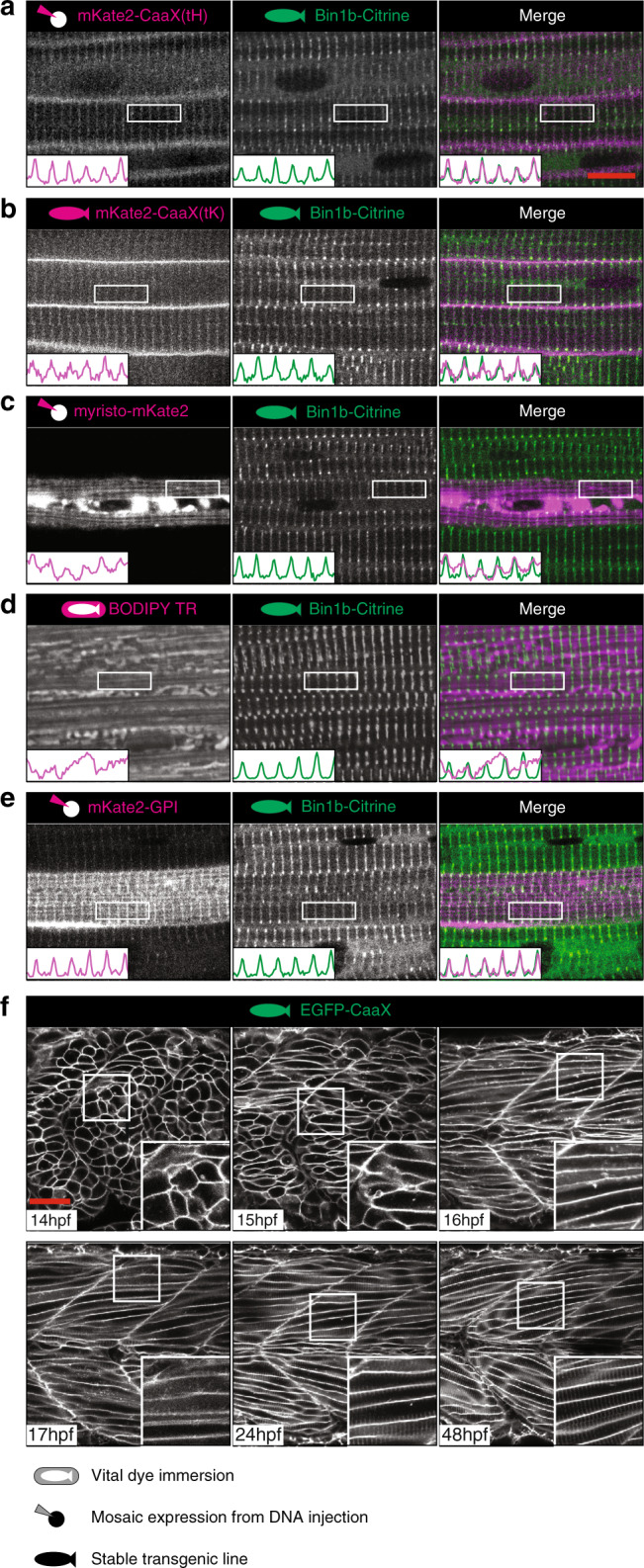
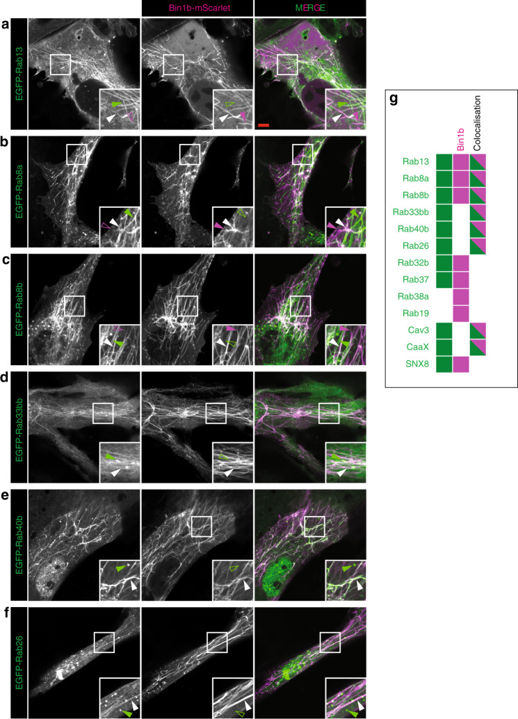
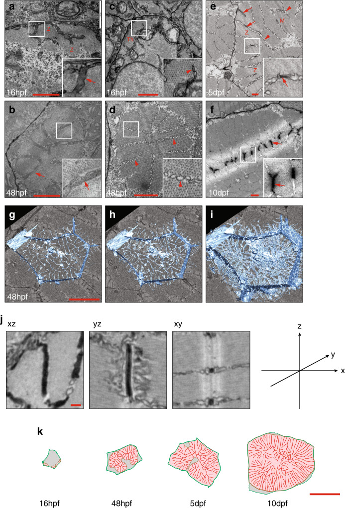
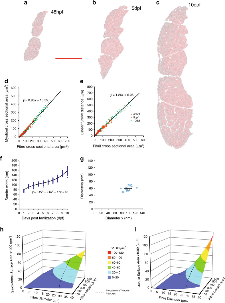
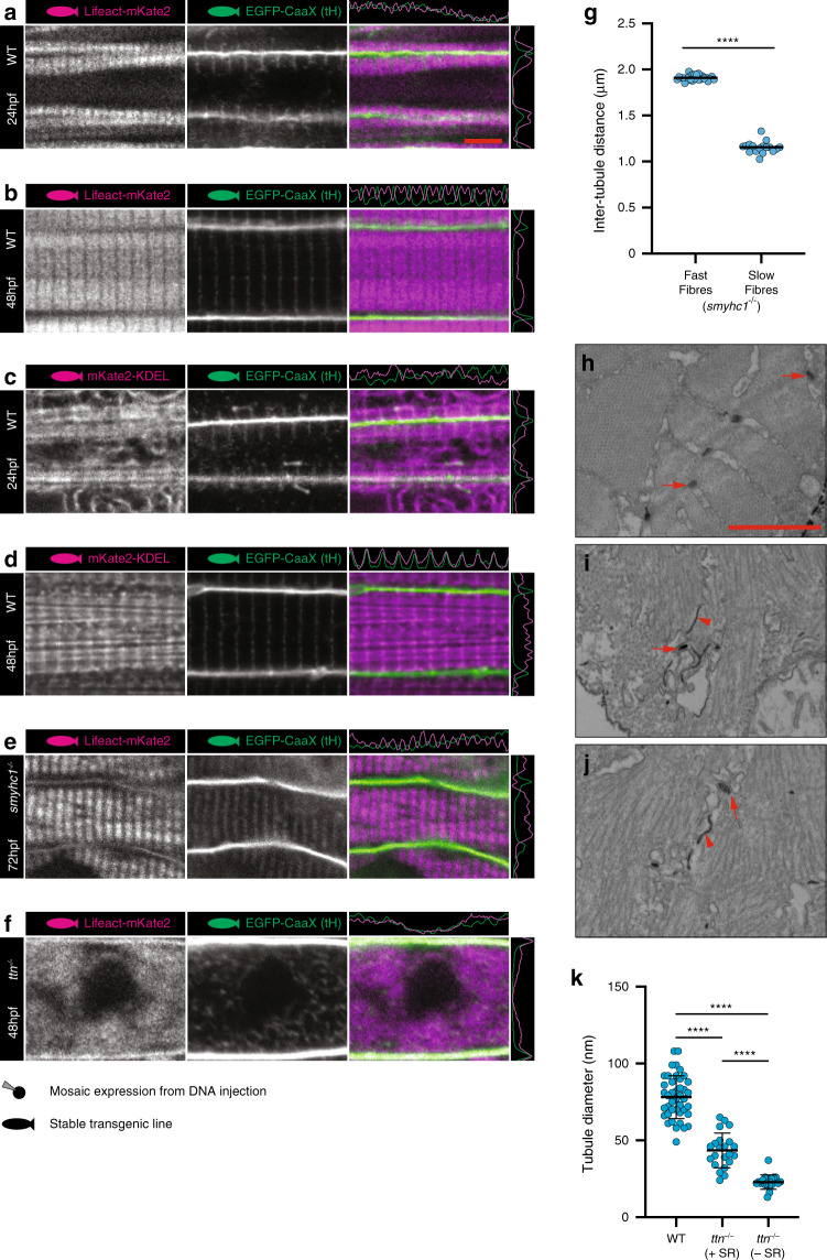
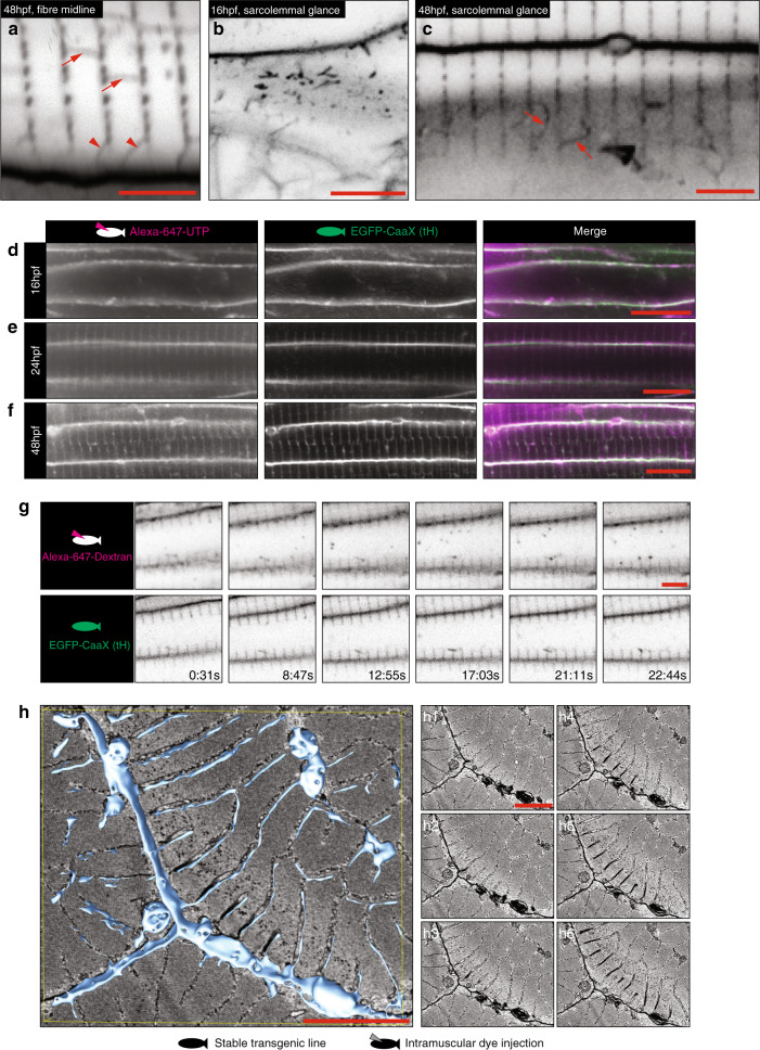
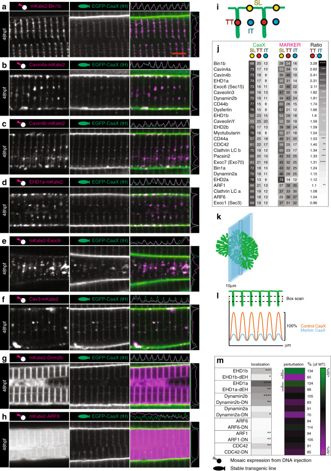
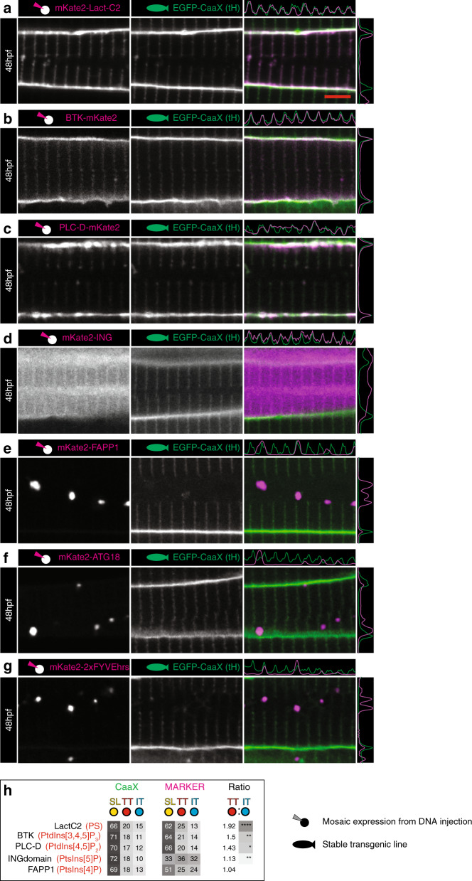
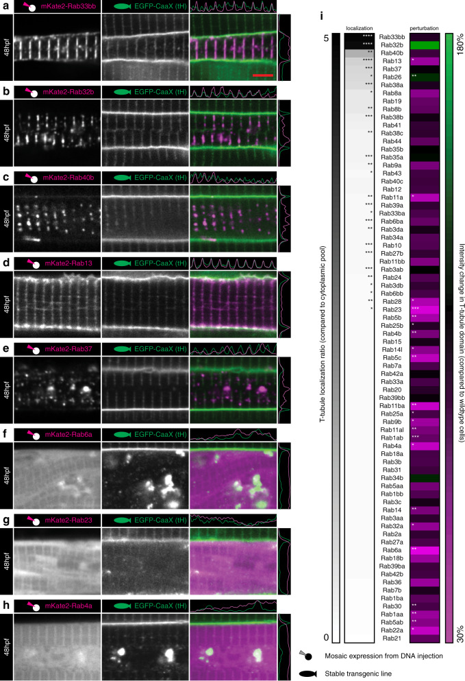
The skeletal muscle T-tubule is a specialized membrane domain essential for coordinated muscle contraction. However, in the absence of genetically tractable systems the mechanisms involved in T-tubule formation are unknown. Here, we use the optically transparent and genetically tractable zebrafish system to probe T-tubule development in vivo. By combining live imaging of transgenic markers with three-dimensional electron microscopy, we derive a four-dimensional quantitative model for T-tubule formation. To elucidate the mechanisms involved in T-tubule formation in vivo, we develop a quantitative screen for proteins that associate with and modulate early T-tubule formation, including an overexpression screen of the entire zebrafish Rab protein family. We propose an endocytic capture model involving firstly, formation of dynamic endocytic tubules at transient nucleation sites on the sarcolemma, secondly, stabilization by myofibrils/sarcoplasmic reticulum and finally, delivery of membrane from the recycling endosome and Golgi complex.
骨骼肌 T 管是一个专门的膜结构域,对于协调肌肉收缩至关重要。然而,在缺乏可遗传的方法的情况下,T 管形成的机制尚不清楚。在这里,我们使用透明且可遗传的斑马鱼系统来探测体内 T 管的发育。通过将转基因标记的活体成像与三维电子显微镜相结合,我们推导出了一个用于 T 管形成的四维定量模型。为了阐明体内 T 管形成的机制,我们开发了一种定量筛选与早期 T 管形成相关并调节其形成的蛋白质的方法,包括对整个斑马鱼 Rab 蛋白家族的过表达筛选。我们提出了一个内吞捕获模型,其中包括:首先,在肌膜上的瞬时成核位点形成动态内吞小管;其次,通过肌原纤维/肌浆网稳定;最后,从再循环内体和高尔基体中输送膜。