Department of Ophthalmology, University of Cincinnati College of Medicine, Cincinnati, OH, USA.
Graefes Arch Clin Exp Ophthalmol. 2021 Jan;259(1):191-196. doi: 10.1007/s00417-020-04851-0. Epub 2020 Jul 25.
To define, describe, and illustrate a previously unreported category of discrete melanotic choroidal melanocytic lesion.
Prospective ophthalmoscopic study of the ocular fundi of 79 light-skinned persons 50 years of age or older not referred for any evident fundus lesion, with detection of all evident discrete melanotic choroidal lesions > 0.3 mm in largest basal diameter.
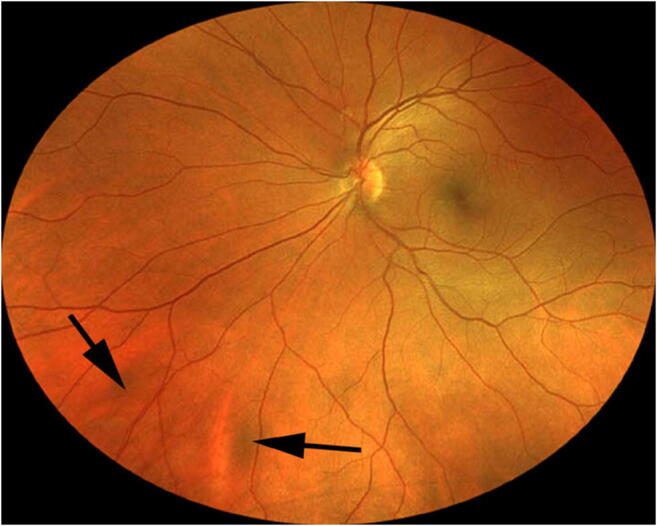
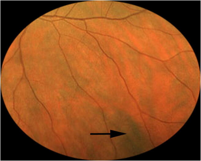
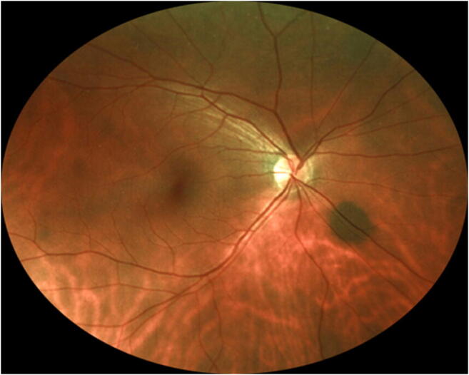
One or more discrete dark-brown to gray choroidal lesions > 0.3 mm in largest basal diameter were detected in 27 of the 79 evaluated subjects (34.2%). All but four of the detected lesions were "flat" by both ophthalmoscopy and ultrasonography. A single flat lesion was present in one eye of 14 subjects whose fellow eye was normal, 2 or more flat lesions were evident in one eye of 5 subjects whose other eye was normal, and one or more lesions were evident in both eyes of 6 subjects.
While some of the discrete small, flat melanocytic choroidal lesions detected in this study might have been choroidal nevi, the author hypothesizes that an indeterminate proportion of them may have been focal aggregates of normal or near normal uveal melanocytes (FANNUMs).
定义、描述和说明一种以前未报告的离散性黑色素性脉络膜黑色素细胞病变类别。
对 79 名年龄在 50 岁或以上、无明显眼底病变但因其他原因就诊的浅色人种的眼部进行前瞻性眼底检查,检查所有明显的最大基底直径大于 0.3 毫米的离散性黑色素性脉络膜病变。
在 79 名受评估者中,有 27 名(34.2%)发现了一个或多个最大基底直径大于 0.3 毫米的离散性深褐色至灰色脉络膜病变。除了四个病变之外,所有病变在眼底镜和超声检查下均为“扁平”。14 名受检者的一只眼存在单个扁平病变,而另一只眼正常;5 名受检者的一只眼存在两个或更多的扁平病变,而另一只眼正常;6 名受检者的双眼均存在一个或多个病变。
虽然本研究中发现的一些离散性小的扁平黑色素性脉络膜病变可能是脉络膜痣,但作者推测,其中一部分可能是正常或接近正常葡萄膜黑色素细胞的局灶性聚集(FANNUMs)。