Lee Joon Seok, Kim Jong Seong, Lee Jeong Woo, Choi Kang Young, Yang Jung Dug, Chung Ho Yun, Cho Byung Chae
Department of Plastic and Reconstructive Surgery, School of Medicine, Kyungpook National University, Daegu, Korea.
Arch Plast Surg. 2020 Jul;47(4):317-323. doi: 10.5999/aps.2018.01165. Epub 2020 Jul 15.
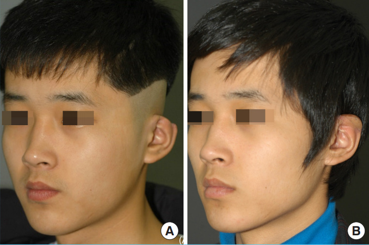
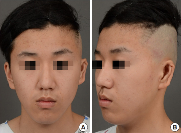
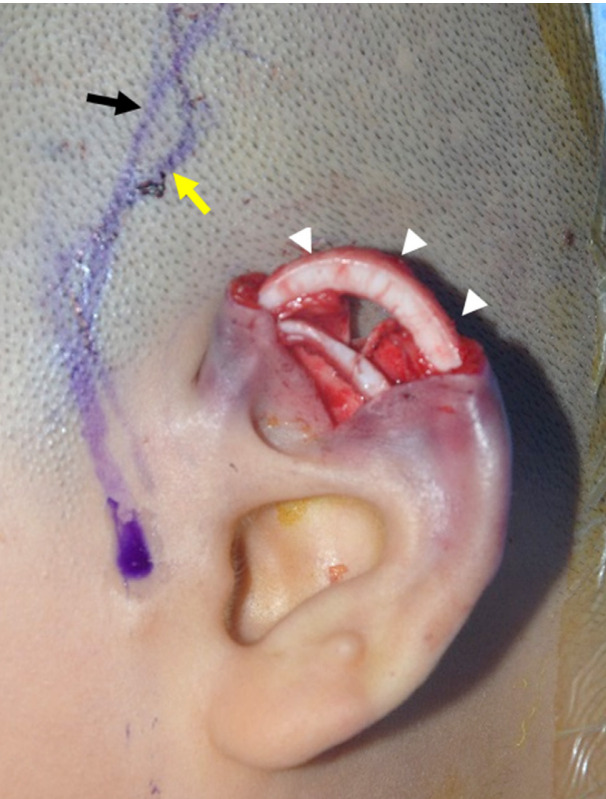
Microtia with constricted features is characterized by a short helical length of variable severity, upper antihelical or scaphal deficiency, and a downfolded upper ear. No consensus has been reached regarding the most appropriate surgical method for this condition. In this study, we aimed to introduce a simple and safe surgical method for the correction or reconstruction of upper helix ear deformities.
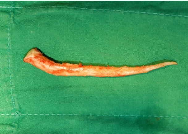
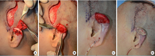
Between February 2011 and June 2014, eight patients with microtia with constricted upper helix ear deformity underwent reconstruction of the ear deformity. The upper ear helical framework was constructed by carving and curving the eighth rib cartilage harvested from the ipsilateral chest wall, covering this cartilage with a superficial temporal fascial flap, and adjusting the skin graft to align with the ear contour. To evaluate their satisfaction, patients were asked to complete a questionnaire regarding ear shape, symmetry, position, color, and overall outcome scored on a 5-point scale at 12 months postoperatively.
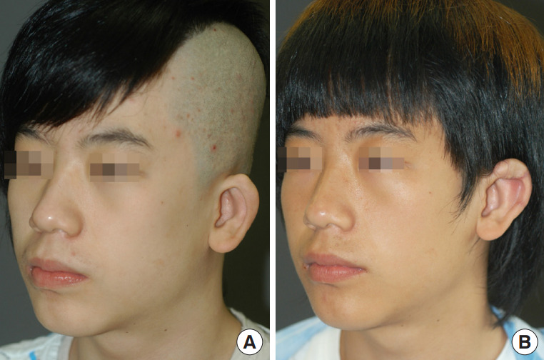
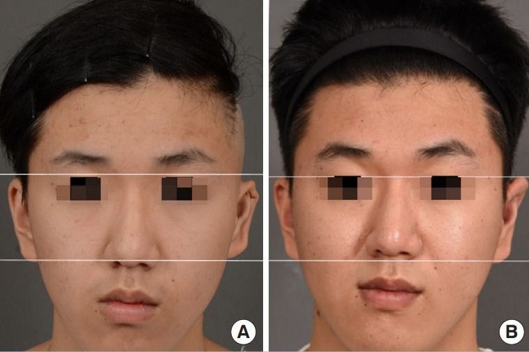
None of the patients experienced severe complications in the reconstructed ear. The preoperative and postoperative vertical ear length ratios were 0.88 and 1.02, respectively. And the mean patient satisfaction scores for shape, symmetry, position, color, and overall outcome were 4.2, 4.5, 4.7, 4.4, and 4.6 out of 5 points, respectively. All patients expressed a high level of satisfaction at 12 months postoperatively.
Our technique provides a good alternative method for the reconstruction of moderate constricted upper helix ear deformities in patients who meet the surgical indications with satisfactory outcomes and few complications.
具有狭窄特征的小耳畸形的特点是螺旋长度短,严重程度不一,上耳轮脚或耳舟缺失,以及上耳向下折叠。对于这种情况最合适的手术方法尚未达成共识。在本研究中,我们旨在介绍一种简单安全的手术方法,用于矫正或重建上耳轮畸形。
2011年2月至2014年6月,8例患有上耳轮狭窄畸形的小耳畸形患者接受了耳畸形重建手术。通过雕刻和弯曲从同侧胸壁获取的第8肋软骨构建上耳轮框架,用颞浅筋膜瓣覆盖该软骨,并调整皮肤移植以使其与耳部轮廓对齐。为了评估他们的满意度,要求患者在术后12个月完成一份关于耳朵形状、对称性、位置、颜色和总体结果的问卷,评分采用5分制。
所有患者重建耳均未出现严重并发症。术前和术后垂直耳长比分别为0.88和1.02。患者对形状、对称性、位置、颜色和总体结果的平均满意度评分分别为4.2分、4.5分、4.7分、4.4分和4.6分(满分5分)。所有患者在术后12个月均表示高度满意。
我们的技术为符合手术指征的患者重建中度狭窄的上耳轮畸形提供了一种良好的替代方法,效果满意,并发症少。