Wang Zhanwen, Li Hong, Long Zeling, Lin Subin, Thoreson Andrew R, Moran Steven L, Gingery Anne, Amadio Peter C, Steinmann Scott P, Zhao Chunfeng
Department of Orthopedic Surgery, Mayo Clinic, Rochester, Minnesota, USA.
Department of Sports Medicine, Xiangya Hospital, Central South University, Changsha, China.
Bone Joint Res. 2020 Jul 23;9(6):285-292. doi: 10.1302/2046-3758.96.BJR-2019-0196.R1. eCollection 2020 Jun.
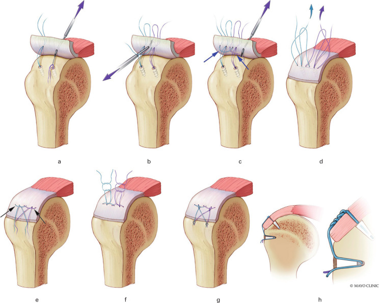
Many biomechanical studies have shown that the weakest biomechanical point of a rotator cuff repair is the suture-tendon interface at the medial row. We developed a novel double rip-stop (DRS) technique to enhance the strength at the medial row for rotator cuff repair. The objective of this study was to evaluate the biomechanical properties of the DRS technique with the conventional suture-bridge (SB) technique and to evaluate the biomechanical performance of the DRS technique with medial row knots.
A total of 24 fresh-frozen porcine shoulders were used. The infraspinatus tendons were sharply dissected and randomly repaired by one of three techniques: SB repair (SB group), DRS repair (DRS group), and DRS with medial row knots repair (DRSK group). Specimens were tested to failure. In addition, 3 mm gap formation was measured and ultimate failure load, stiffness, and failure modes were recorded.
The mean load to create a 3 mm gap formation in the DRSK and DRS groups was significantly higher than in the SB group. The DRSK group had the highest load to failure with a mean ultimate failure load of 395.0 N (SD 56.8) compared to the SB and DRS groups, which recorded 147.1 N (SD 34.3) and 285.9 N (SD 89.8), respectively (p < 0.001 for both). The DRS group showed a significantly higher mean failure load than the SB group (p = 0.006). Both the DRS and DRSK groups showed significantly higher mean stiffness than the SB group.
The biomechanical properties of the DRS technique were significantly improved compared to the SB technique. The DRS technique with medial row knots showed superior biomechanical performance than the DRS technique alone.
许多生物力学研究表明,肩袖修复最薄弱的生物力学点是内侧排的缝线-肌腱界面。我们开发了一种新型双止裂(DRS)技术,以增强肩袖修复内侧排的强度。本研究的目的是评估DRS技术与传统缝线桥接(SB)技术的生物力学性能,并评估带内侧排结的DRS技术的生物力学性能。
共使用24个新鲜冷冻猪肩。将冈下肌腱锐性解剖,随机采用三种技术之一进行修复:SB修复(SB组)、DRS修复(DRS组)和带内侧排结的DRS修复(DRSK组)。对标本进行直至失效的测试。此外,测量3mm间隙形成情况,并记录极限失效载荷、刚度和失效模式。
DRSK组和DRS组产生3mm间隙形成的平均载荷显著高于SB组。DRSK组的失效载荷最高,平均极限失效载荷为395.0N(标准差56.8),而SB组和DRS组分别为147.1N(标准差34.3)和285.9N(标准差89.8)(两者p均<0.001)。DRS组的平均失效载荷显著高于SB组(p=0.006)。DRS组和DRSK组的平均刚度均显著高于SB组。
与SB技术相比,DRS技术的生物力学性能有显著改善。带内侧排结的DRS技术比单独的DRS技术表现出更优异的生物力学性能。