• 文献检索
  • 文档翻译
  • 深度研究
  • 学术资讯
  • Suppr Zotero 插件Zotero 插件
  • 邀请有礼
  • 套餐&价格
  • 历史记录
应用&插件
Suppr Zotero 插件Zotero 插件浏览器插件Mac 客户端Windows 客户端微信小程序
定价
高级版会员购买积分包购买API积分包
服务
文献检索文档翻译深度研究API 文档MCP 服务
关于我们
关于 Suppr公司介绍联系我们用户协议隐私条款
关注我们

Suppr 超能文献

核心技术专利:CN118964589B侵权必究
粤ICP备2023148730 号-1Suppr @ 2026

文献检索

告别复杂PubMed语法,用中文像聊天一样搜索,搜遍4000万医学文献。AI智能推荐,让科研检索更轻松。

立即免费搜索

文件翻译

保留排版,准确专业,支持PDF/Word/PPT等文件格式,支持 12+语言互译。

免费翻译文档

深度研究

AI帮你快速写综述,25分钟生成高质量综述,智能提取关键信息,辅助科研写作。

立即免费体验

利用中子衍射和中子计算机层析成像技术分析珊瑚骨骼的结构。

The structure of scleractinian coral skeleton analyzed by neutron diffraction and neutron computed tomography.

机构信息

Frank Laboratory for Neutron Physics, Joint Institute for Nuclear Research, 6, Joliot Curie str, 141980, Dubna, Russian Federation.

Department of Structure of Matter, Earth and Atmospheric Physics and Astrophysics, Faculty of Physics, University of Bucharest, 405, Atomistilor str., 077125, Magurele, Ilfov, Romania.

出版信息

Sci Rep. 2020 Jul 30;10(1):12869. doi: 10.1038/s41598-020-69859-2.

DOI:10.1038/s41598-020-69859-2
PMID:32733083
原文链接:https://pmc.ncbi.nlm.nih.gov/articles/PMC7392896/
Abstract

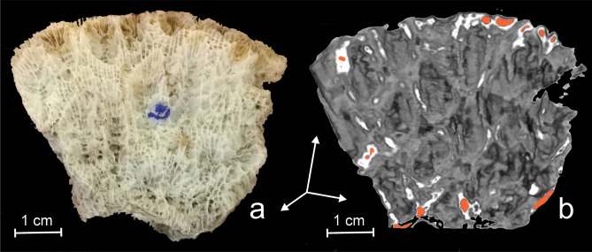
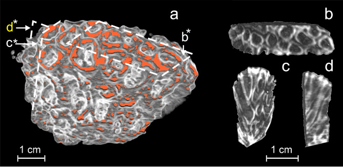
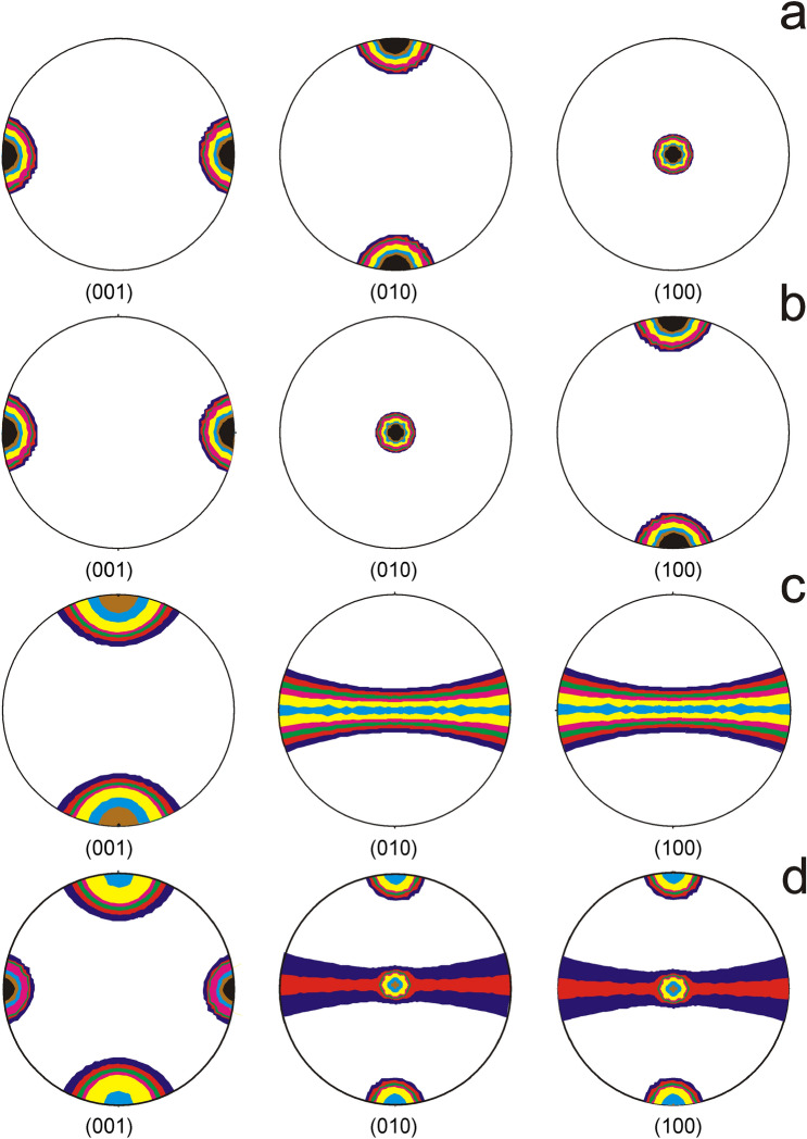
Two analytical methods based on the neutrons high penetrability, i.e. neutron diffraction (ND) and neutron computed tomography (NCT) were used to investigate the structure of the aragonitic skeleton of an exemplar/sample of Dipastraea pallida (Dana 1846), a modern hermatypic coral. ND was used to reconstruct the orientation distribution function (ODF) of the crystalline fibrils which compose the coral skeleton. Accordingly, 684 ND spectra were analyzed using the Rietveld method. The result confirmed the aragonite as the sole mineral component of coral skeleton, allowing to recalculate the ODF of aragonite fibrils and to represent it by means of (100), (010) and (001) crystallographic planes pole figures (PF). Experimental PF showed a remarkable similarity with PF recalculated by considering that all aragonite fibrils are oriented either along the growth axis of polyp cups or perpendicular to this direction. This result confirmed the previous observations based on optical microscopy, proving at the same time the availability of ND for such types of investigations. In turn, NCT evidenced the individual polyp cups, their interlocked 3D rigid porous structure as well as a periodic variation of density which could be attributed to a seasonal influence of the marine environment. Different from the classical X-ray computed tomography, the NCT, in view of neutron high cross-section for hydrogen, demonstrated the presence of a small amount of organic matter, otherwise transparent for X- and gamma rays.

摘要

两种基于中子高穿透性的分析方法,即中子衍射(ND)和中子计算机断层扫描(NCT),被用于研究现代四射珊瑚 Dipastraea pallida(Dana 1846)的方解石骨骼结构。ND 用于重建组成珊瑚骨骼的晶体原纤的取向分布函数(ODF)。相应地,使用 Rietveld 方法分析了 684 个 ND 光谱。结果证实了方解石是珊瑚骨骼的唯一矿物成分,从而可以重新计算方解石原纤的 ODF,并通过(100)、(010)和(001)晶面极图(PF)来表示。实验 PF 与考虑所有方解石原纤沿珊瑚杯生长轴或垂直于该方向取向时重新计算的 PF 具有显著相似性。这一结果证实了先前基于光学显微镜的观察结果,同时证明了 ND 适用于此类研究。反过来,NCT 证明了单个珊瑚杯、它们相互锁定的 3D 刚性多孔结构以及密度的周期性变化,这可能归因于海洋环境的季节性影响。与经典的 X 射线计算机断层扫描不同,鉴于中子对氢的高截面,NCT 显示出存在少量的有机物,否则对 X 射线和伽马射线是透明的。

https://cdn.ncbi.nlm.nih.gov/pmc/blobs/f216/7392896/ffff579e7dc7/41598_2020_69859_Fig7_HTML.jpg
https://cdn.ncbi.nlm.nih.gov/pmc/blobs/f216/7392896/156e9b3c610f/41598_2020_69859_Fig1_HTML.jpg
https://cdn.ncbi.nlm.nih.gov/pmc/blobs/f216/7392896/ad38dde066ae/41598_2020_69859_Fig2_HTML.jpg
https://cdn.ncbi.nlm.nih.gov/pmc/blobs/f216/7392896/e759116c853f/41598_2020_69859_Fig3_HTML.jpg
https://cdn.ncbi.nlm.nih.gov/pmc/blobs/f216/7392896/903173f59455/41598_2020_69859_Fig4_HTML.jpg
https://cdn.ncbi.nlm.nih.gov/pmc/blobs/f216/7392896/ffe5d76e286f/41598_2020_69859_Fig5_HTML.jpg
https://cdn.ncbi.nlm.nih.gov/pmc/blobs/f216/7392896/a9aecbca9a74/41598_2020_69859_Fig6_HTML.jpg
https://cdn.ncbi.nlm.nih.gov/pmc/blobs/f216/7392896/ffff579e7dc7/41598_2020_69859_Fig7_HTML.jpg

相似文献

1
The structure of scleractinian coral skeleton analyzed by neutron diffraction and neutron computed tomography.利用中子衍射和中子计算机层析成像技术分析珊瑚骨骼的结构。
Sci Rep. 2020 Jul 30;10(1):12869. doi: 10.1038/s41598-020-69859-2.
2
A modern scleractinian coral with a two-component calcite-aragonite skeleton.具有两部分方解石-霰石骨骼的现代珊瑚。
Proc Natl Acad Sci U S A. 2021 Jan 19;118(3). doi: 10.1073/pnas.2013316117. Epub 2020 Dec 15.
3
Biotic control of skeletal growth by scleractinian corals in aragonite-calcite seas.文石-方解石海中生硬珊瑚对骨骼生长的生物控制。
PLoS One. 2014 Mar 7;9(3):e91021. doi: 10.1371/journal.pone.0091021. eCollection 2014.
4
The two-step mode of growth in the scleractinian coral skeletons from the micrometre to the overall scale.石珊瑚骨骼从微米级到整体尺度的两步生长模式。
J Struct Biol. 2005 Jun;150(3):319-31. doi: 10.1016/j.jsb.2005.03.004. Epub 2005 Mar 31.
5
Needle-like grains across growth lines in the coral skeleton of Porites lobata.针状颗粒穿过鹿角杯形珊瑚骨骼的生长线。
J Struct Biol. 2012 Dec;180(3):389-93. doi: 10.1016/j.jsb.2012.09.009. Epub 2012 Oct 4.
6
Corals concentrate dissolved inorganic carbon to facilitate calcification.珊瑚浓缩溶解的无机碳以促进钙化。
Nat Commun. 2014 Dec 22;5:5741. doi: 10.1038/ncomms6741.
7
Biomineralization in newly settled recruits of the scleractinian coral Pocillopora damicornis.新附着的鹿角杯形珊瑚幼体中的生物矿化作用。
J Morphol. 2014 Dec;275(12):1349-65. doi: 10.1002/jmor.20307. Epub 2014 Jun 25.
8
Coral biomineralization: A focus on intra-skeletal organic matrix and calcification.珊瑚生物矿化:聚焦骨骼内有机基质与钙化
Semin Cell Dev Biol. 2015 Oct;46:17-26. doi: 10.1016/j.semcdb.2015.09.005. Epub 2015 Sep 5.
9
Aragonite precipitation by "proto-polyps" in coral cell cultures.珊瑚细胞培养中的“原息肉”对文石的沉淀作用。
PLoS One. 2012;7(4):e35049. doi: 10.1371/journal.pone.0035049. Epub 2012 Apr 13.
10
Comparative Proteomics of Octocoral and Scleractinian Skeletomes and the Evolution of Coral Calcification.八放珊瑚和石珊瑚骨骼的比较蛋白质组学与珊瑚钙化的演化。
Genome Biol Evol. 2020 Sep 1;12(9):1623-1635. doi: 10.1093/gbe/evaa162.

引用本文的文献

1
Polyp-Canal Reconstruction Reveals Evolution Toward Complexity in Corals.息肉-管道重建揭示了珊瑚向复杂性的进化。
Research (Wash D C). 2023 Jun 6;6:0166. doi: 10.34133/research.0166. eCollection 2023.
2
Micro-CT reconstruction reveals the colony pattern regulations of four dominant reef-building corals.微计算机断层扫描重建揭示了四种主要造礁珊瑚的群体模式规律。
Ecol Evol. 2021 Nov 4;11(22):16266-16279. doi: 10.1002/ece3.8308. eCollection 2021 Nov.