Department of Radiology, Poznań University of Medical Sciences, Poznań, Poland.
Fast-Radiology, Poland.
PLoS One. 2020 Jul 31;15(7):e0237092. doi: 10.1371/journal.pone.0237092. eCollection 2020.
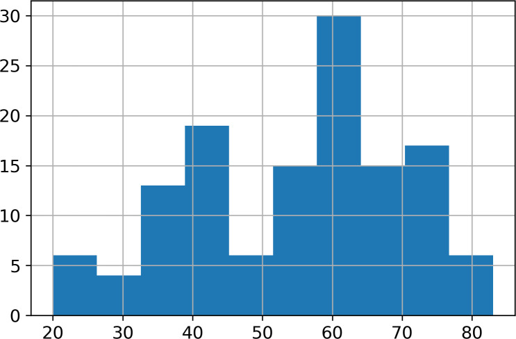
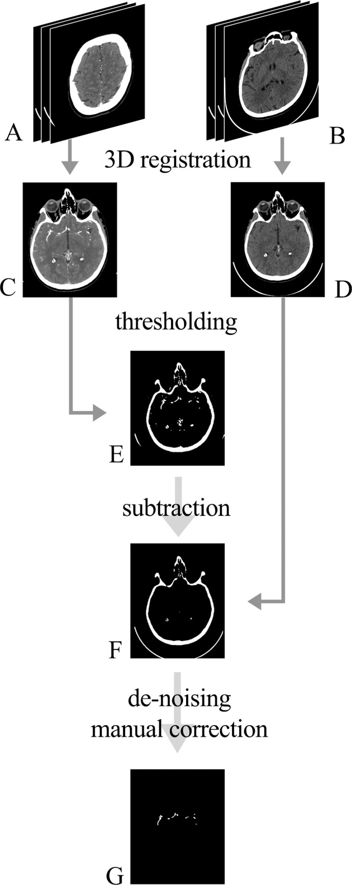
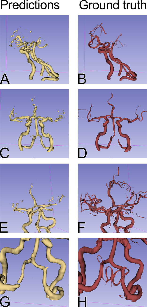
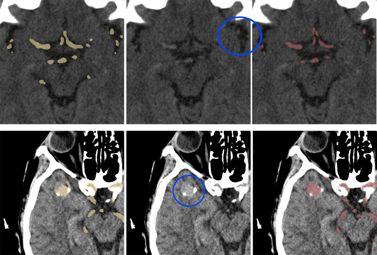
Cerebral computed tomography angiography is a widely available imaging technique that helps in the diagnosis of vascular pathologies. Contrast administration is needed to accurately assess the arteries. On non-contrast computed tomography, arteries are hardly distinguishable from the brain tissue, therefore, radiologists do not consider this imaging modality appropriate for the evaluation of vascular pathologies. There are known contraindications to administering iodinated contrast media, and in these cases, the patient has to undergo another examination to visualize cerebral arteries, such as magnetic resonance angiography. Deep learning for image segmentation has proven to perform well on medical data for a variety of tasks. The aim of this research was to apply deep learning methods to segment cerebral arteries on non-contrast computed tomography scans and consequently, generate angiographies without the need for contrast administration. The dataset for this research included 131 patients who underwent brain non-contrast computed tomography directly followed by computed tomography with contrast administration. Then, the segmentations of arteries were generated and aligned with non-contrast computed tomography scans. A deep learning model based on the U-net architecture was trained to perform the segmentation of blood vessels on non-contrast computed tomography. An evaluation was performed on separate test data, as well as using cross-validation, reaching Dice coefficients of 0.638 and 0.673, respectively. This study proves that deep learning methods can be leveraged to quickly solve problems that are difficult and time-consuming for a human observer, therefore providing physicians with additional information on the patient. To encourage the further development of similar tools, all code used for this research is publicly available.
计算机断层血管造影术是一种广泛应用的成像技术,有助于诊断血管病变。需要进行造影剂给药才能准确评估动脉。在非增强计算机断层扫描中,动脉与脑组织几乎难以区分,因此放射科医生认为这种成像方式不适合评估血管病变。存在碘造影剂给药的已知禁忌症,在这些情况下,患者必须进行另一种检查以可视化脑动脉,例如磁共振血管造影。图像分割的深度学习已被证明在各种任务的医学数据上表现良好。本研究的目的是应用深度学习方法对非增强计算机断层扫描上的脑动脉进行分割,从而无需造影剂给药即可生成血管造影。本研究的数据集中包括 131 名患者,他们在直接进行脑非增强计算机断层扫描后紧接着进行了计算机断层扫描加造影。然后,生成动脉的分割并与非增强计算机断层扫描对齐。基于 U-net 架构的深度学习模型被训练用于执行非增强计算机断层扫描上的血管分割。在单独的测试数据上进行了评估,以及使用交叉验证,分别达到了 0.638 和 0.673 的 Dice 系数。这项研究证明,深度学习方法可以被利用来快速解决对人类观察者来说困难和耗时的问题,从而为医生提供有关患者的额外信息。为了鼓励类似工具的进一步发展,本研究中使用的所有代码都是公开的。