Immunohematology and Blood Transfusion, Leiden University Medical Center, Leiden, Netherlands.
Institute for Immunity, Transplantation and Infection, Stanford University, Stanford, CA, United States.
Front Immunol. 2020 Jul 16;11:1466. doi: 10.3389/fimmu.2020.01466. eCollection 2020.
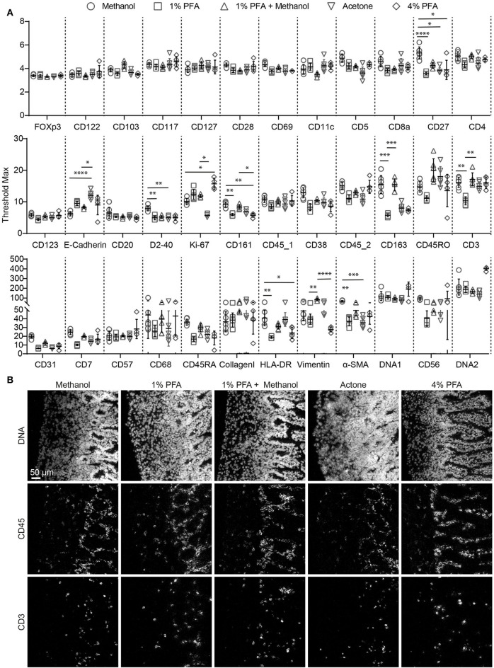
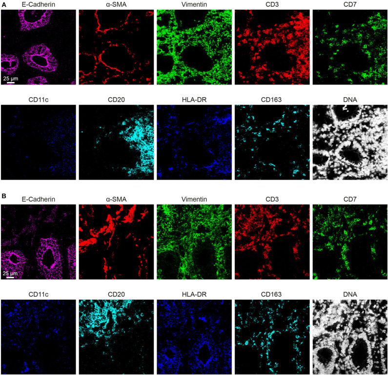
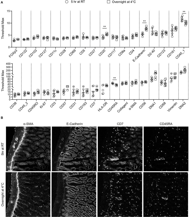
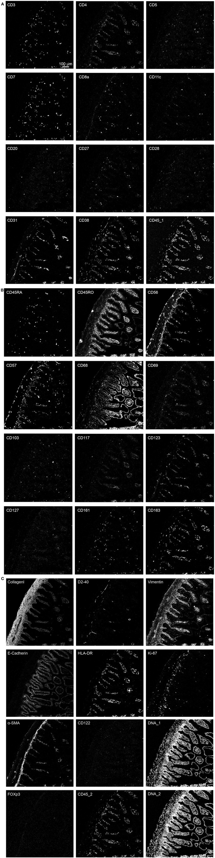
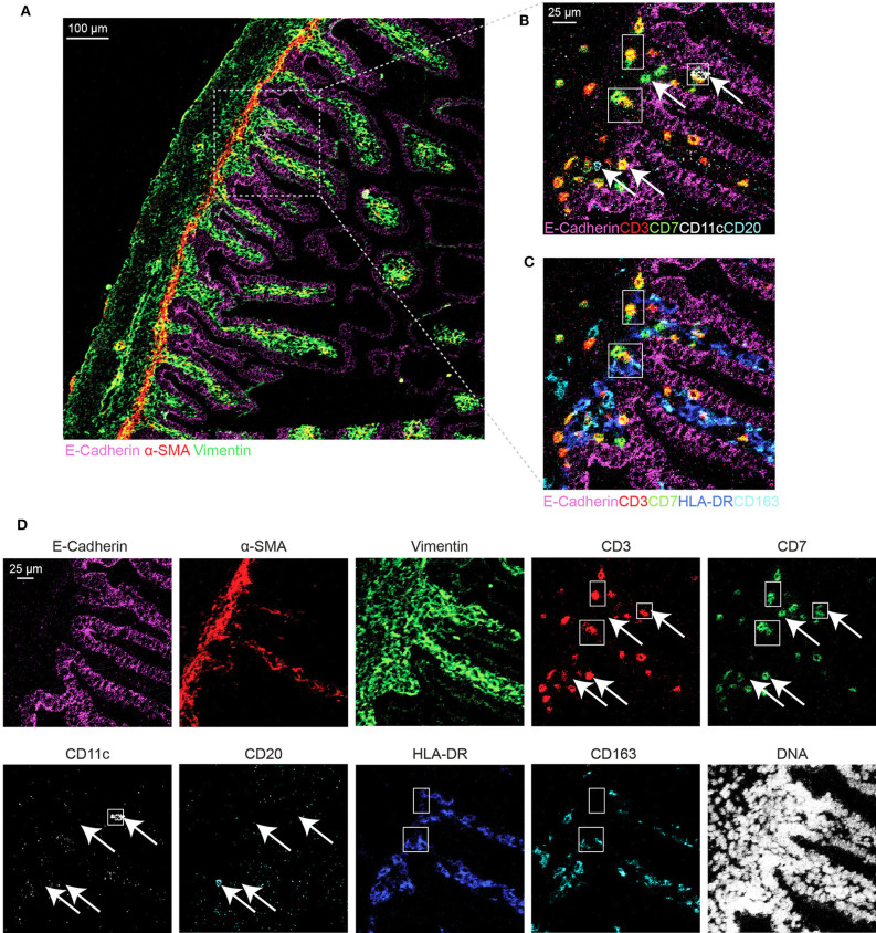
Imaging mass cytometry (IMC) is able to quantify the expression of dozens of markers at sub-cellular resolution on a single tissue section by combining a novel laser ablation system with mass cytometry. As such, it allows us to gain spatial information and antigen quantification , and can be applied to both snap-frozen and formalin-fixed, paraffin-embedded (FFPE) tissue sections. Herein, we have developed and optimized the immunodetection conditions for a 34-antibody panel for use on human snap-frozen tissue sections. For this, we tested the performance of 80 antibodies. Moreover, we compared tissue drying times, fixation procedures and antibody incubation conditions. We observed that variations in the drying times of tissue sections had little impact on the quality of the images. Fixation with methanol for 5 min at -20°C or 1% paraformaldehyde (PFA) for 5 min at room temperature followed by methanol for 5 min at -20°C were superior to fixation with acetone or PFA only. Finally, we observed that antibody incubation overnight at 4°C yielded more consistent results as compared to staining at room temperature for 5 h. Finally, we used the optimized method for staining of human fetal and adult intestinal tissue samples. We present the tissue architecture and spatial distribution of the stromal cells and immune cells in these samples visualizing blood vessels, the epithelium and lamina propria based on the expression of α-smooth muscle actin (α-SMA), E-Cadherin and Vimentin, while simultaneously revealing the colocalization of T cells, innate lymphoid cells (ILCs), and various myeloid cell subsets in the lamina propria of the human fetal intestine. We expect that this work can aid the scientific community who wish to improve IMC data quality.
成像质谱流式细胞术 (IMC) 通过将新型激光烧蚀系统与质谱流式细胞术相结合,能够在单个组织切片上以亚细胞分辨率定量检测数十种标记物的表达。因此,它可以让我们获得空间信息和抗原定量,并可应用于新鲜冷冻和福尔马林固定、石蜡包埋(FFPE)的组织切片。在此,我们针对人新鲜冷冻组织切片开发并优化了包含 34 种抗体的免疫检测条件。为此,我们测试了 80 种抗体的性能。此外,我们还比较了组织干燥时间、固定程序和抗体孵育条件。我们观察到组织切片干燥时间的变化对图像质量的影响很小。在-20°C 下用甲醇固定 5 分钟或在室温下用 1%多聚甲醛(PFA)固定 5 分钟,然后再用甲醇在-20°C 下固定 5 分钟,优于仅用丙酮或 PFA 固定。最后,我们观察到在 4°C 下孵育过夜的抗体比在室温下孵育 5 小时得到的结果更加一致。最后,我们使用优化的方法对人胎儿和成人肠道组织样本进行染色。我们展示了这些样本的组织架构和基质细胞及免疫细胞的空间分布,通过α-平滑肌肌动蛋白(α-SMA)、E-钙黏蛋白和波形蛋白的表达来可视化血管、上皮和固有层,同时揭示了 T 细胞、固有淋巴样细胞(ILCs)和各种髓样细胞亚群在人胎儿肠道固有层中的共定位。我们希望这项工作可以帮助希望提高 IMC 数据质量的科学界。