Division of Gastroenterology, Department of Internal Medicine, Kangbuk Samsung Hospital, Sungkyunkwan University School of Medicine, Seoul, Korea.
Department of Surgery, Kangbuk Samsung Hospital, Sungkyunkwan University School of Medicine, Seoul, Korea.
Korean J Radiol. 2020 Dec;21(12):1355-1366. doi: 10.3348/kjr.2019.0891. Epub 2020 Aug 4.
We aimed to evaluate the diagnostic value and prognostic relevance of FDG positron emission tomography/computed tomography (PET-CT) in extrahepatic cholangiocarcinoma patients.
This study included 234 extrahepatic cholangiocarcinoma patients who underwent FDG PET-CT between June 2008 and February 2016. The diagnostic performance of FDG PEG-CT was compared to that of contrast-enhanced multidetector row CT (MDCT) and MRI. Independent prognosticators for poor survival were also assessed.
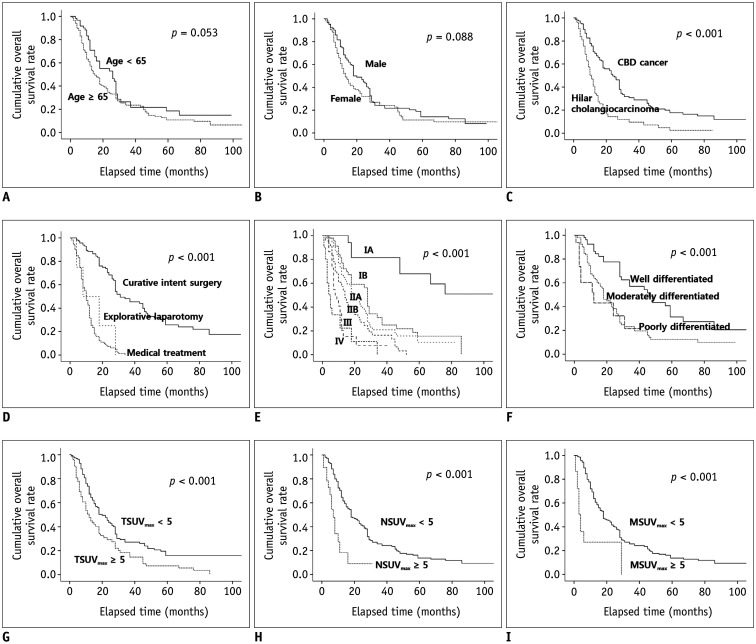
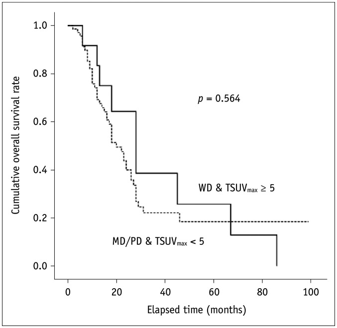
The sensitivity of FDG PET-CT for detecting primary tumor and regional lymph node metastases was lower than that of MDCT or MRI ( < 0.001), whereas the specificity and positive predictive value for detecting regional lymph nodes metastases was significantly better in FDG PET-CT compared to MDCT and MRI (all < 0.001). There was no significant difference in the diagnostic yield of distant metastases detection among three diagnostic imaging techniques. In a multivariate analysis, maximum standardized uptake values (SUV) of the primary tumor (adjusted hazard ratio [HR], 1.75; 95% confidence interval [CI], 1.13-2.69) and of the metastatic lesions ≥ 5 (adjusted HR, 8.10; 95% CI, 1.96-33.5) were independent contributors to poor overall survival in extrahepatic cholangiocarcinoma patients. In a subgroup analysis of 187 patients with periductal infiltrating type of cholangiocarcinoma, an SUV of the primary tumor ≥ 5 was associated with an increased risk of regional lymph node (adjusted odds ratio [OR], 1.60; 95% CI, 0.55-4.63) and distant metastases (adjusted OR, 100.57; 95% CI, 3.94-2567.43) at diagnosis as well as with poor overall survival (adjusted HR, 1.81; 95% CI, 1.04-3.15).
FDG PET-CT showed lower sensitivity for detecting primary tumor and regional lymph node involvement than MDCT and MRI. However, the SUV of primary tumors and metastatic lesions derived from FDG PET-CT could have significant implications for predicting prognoses in extrahepatic cholangiocarcinoma patients.
评估氟代脱氧葡萄糖正电子发射断层扫描/计算机断层扫描(FDG PET-CT)在肝外胆管癌患者中的诊断价值和预后相关性。
本研究纳入了 234 例于 2008 年 6 月至 2016 年 2 月间行 FDG PET-CT 检查的肝外胆管癌患者。将 FDG PET-CT 的诊断性能与对比增强多排螺旋 CT(MDCT)和 MRI 进行比较。还评估了影响不良生存的独立预后因素。
FDG PET-CT 检测原发肿瘤和区域淋巴结转移的敏感性低于 MDCT 或 MRI(均<0.001),而 FDG PET-CT 检测区域淋巴结转移的特异性和阳性预测值明显优于 MDCT 和 MRI(均<0.001)。三种诊断影像学技术在远处转移检测的诊断效能上无显著差异。多因素分析显示,原发肿瘤的最大标准化摄取值(SUV)(调整后的危险比[HR],1.75;95%置信区间[CI],1.13-2.69)和转移病灶 SUV≥5(调整后的 HR,8.10;95%CI,1.96-33.5)是肝外胆管癌患者总体生存不良的独立预后因素。在 187 例胆管壁周围浸润型胆管癌患者的亚组分析中,原发肿瘤 SUV≥5 与区域淋巴结(调整后的优势比[OR],1.60;95%CI,0.55-4.63)和远处转移(调整后的 OR,100.57;95%CI,3.94-2567.43)的诊断风险增加以及总体生存不良(调整后的 HR,1.81;95%CI,1.04-3.15)相关。
FDG PET-CT 检测原发肿瘤和区域淋巴结受累的敏感性低于 MDCT 和 MRI。然而,源自 FDG PET-CT 的原发肿瘤和转移病灶 SUV 对预测肝外胆管癌患者的预后具有重要意义。