Department of Cytology and Gynecological Pathology, Postgraduate Institute of Medical Education and Research, CHANDIGARH, INDIA.
Turk Patoloji Derg. 2021;37(1):84-88. doi: 10.5146/tjpath.2020.01501.
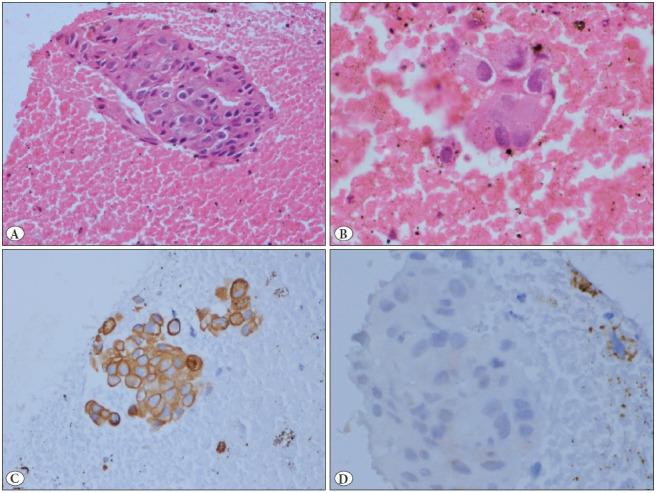
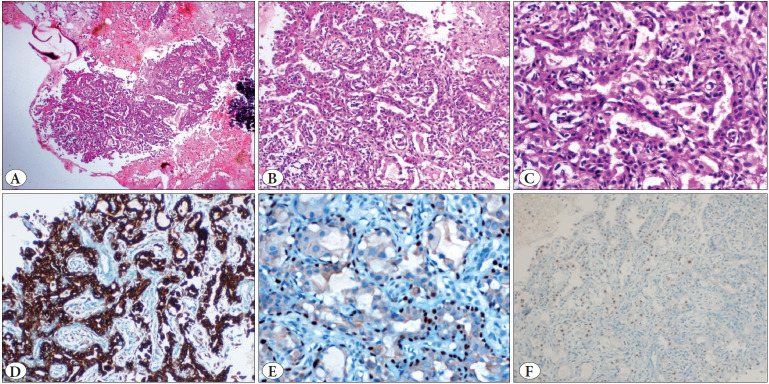
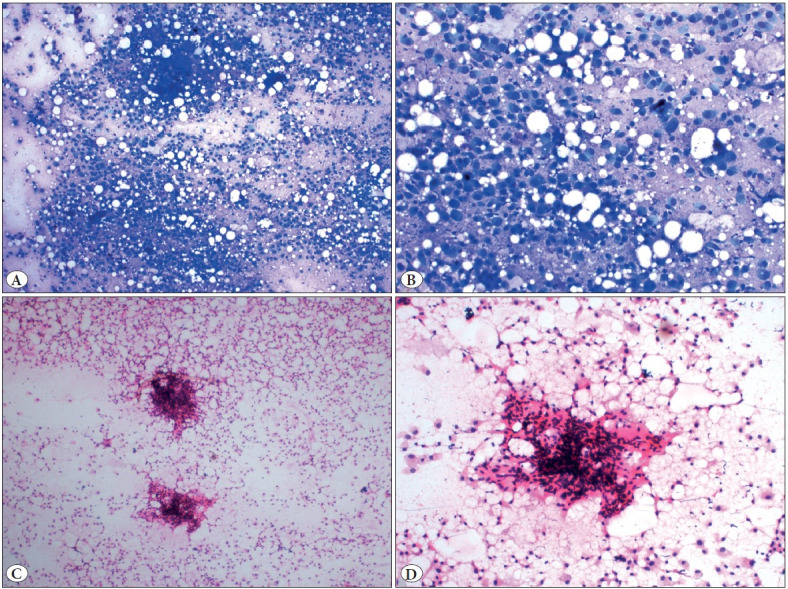
Epithelial-myoepithelial carcinoma (EMC) is a rare, low-grade, malignant salivary neoplasm. Establishing an accurate cytological diagnosis is often challenging owing to its rarity, bland cytologic appearance and variable representation of cell populations in the smears. The diagnostic struggle is more so when the aspiration is from a metastatic site with an unknown primary, as in such cases the list of differential diagnoses expands further. A 58-year-old female presented with a low-back pain from last one month. On examination, she also had a level III, right cervical swelling for the last 20 years. Radiology revealed a lytic lesion in the left acetabulum. She had undergone surgery 35 years ago for a right-sided upper neck swelling, the medical records of which were not available. Fine needle aspiration (FNA) from the cervical swelling was performed. The smears were cellular and showed predominantly dispersed, round to polygonal tumor cells with mild pleomorphism, eccentric nuclei, coarse chromatin, occasional nucleoli and moderate cytoplasm with some showing vacuolations. The cell-block section revealed tumor cells arranged in the form of tubules lined by dual layer of tumor cells without any chondromyxoid stroma. On immunocytochemistry, the luminal cells showed positivity for CK7 (epithelial marker) and the abluminal cells showed positivity for p63 (myoepithelial marker). Based on these features, a final diagnosis of metastatic epithelial-myoepithelial carcinoma was rendered. The present report highlights the characteristic cytomorphological and immunocytochemical features of EMC and reiterates the diagnostic accuracy of FNAC for diagnosis of such challenging cases.
上皮-肌上皮癌(EMC)是一种罕见的低级别恶性涎腺肿瘤。由于其罕见性、温和的细胞学表现以及在涂片上细胞群体的不同表现,因此准确进行细胞学诊断常常具有挑战性。当抽吸物来自未知原发灶的转移部位时,诊断难度更大,因为在这种情况下,鉴别诊断的范围进一步扩大。一名 58 岁女性因腰痛就诊,已持续一个月。体格检查发现她的右侧颈部有一个 III 级肿大,已经持续 20 年。影像学检查显示左髋臼有一个溶骨性病变。她 35 年前曾因右侧颈部肿胀接受过手术,但无法获得相关病历记录。对颈部肿胀进行了细针抽吸(FNA)。涂片显示细胞丰富,主要为分散的圆形至多边形肿瘤细胞,轻度多形性,偏心核,粗糙染色质,偶尔有核仁,中等量细胞质,部分细胞有空泡。细胞块切片显示肿瘤细胞呈双层排列的管状结构,没有软骨黏液样基质。免疫细胞化学染色显示,腔细胞对 CK7(上皮标志物)呈阳性,腔旁细胞对 p63(肌上皮标志物)呈阳性。根据这些特征,最终诊断为转移性上皮-肌上皮癌。本报告强调了 EMC 的特征性细胞学和免疫细胞化学特征,并再次强调了细针抽吸在诊断此类具有挑战性病例中的准确性。