Xu Xiaojuan, Lin Sisi, Yang Yanhua, Gong Xiaohui, Tong Jianhua, Li Kun, Li Yongyu
Department of Pathology and Pathophysiology, Tongji University School of Medicine, Shanghai 200092, P.R. China.
Key Laboratory of Arrhythmias of the Ministry of Education of China, Shanghai East Hospital, Shanghai 200120, P.R. China.
Exp Ther Med. 2020 Sep;20(3):1987-1994. doi: 10.3892/etm.2020.8946. Epub 2020 Jun 25.
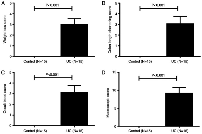
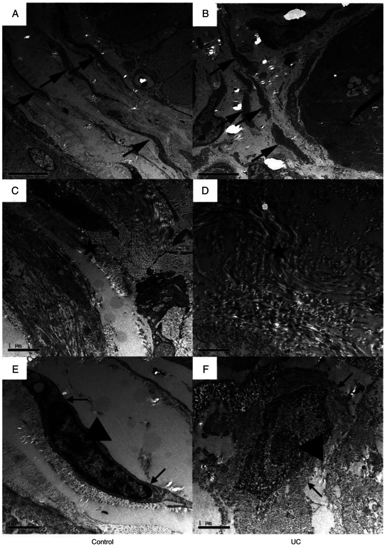
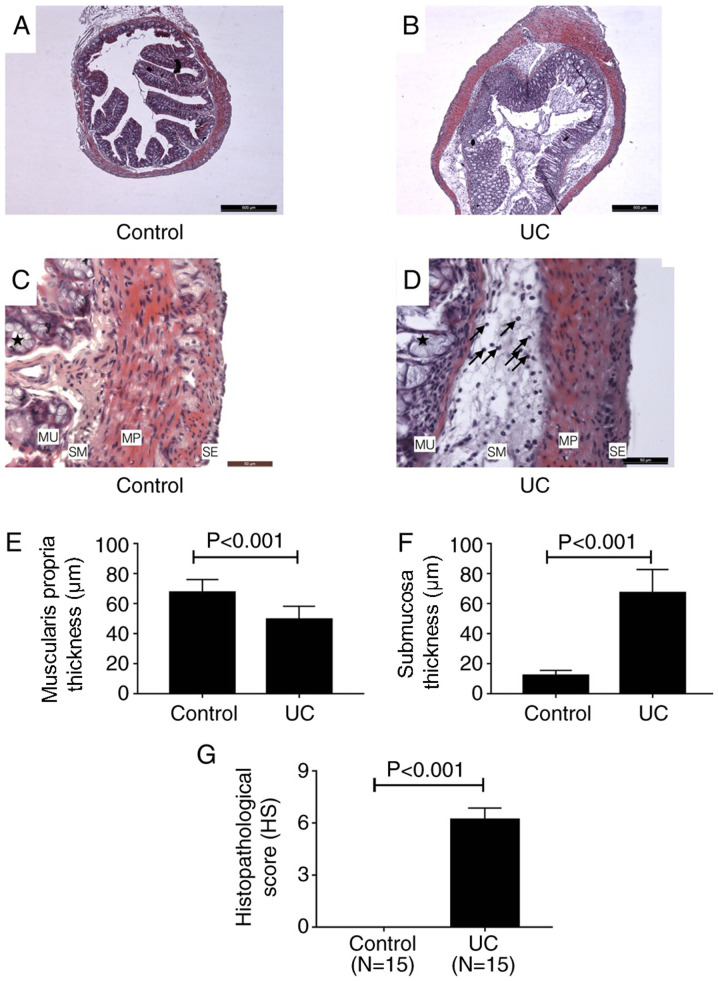
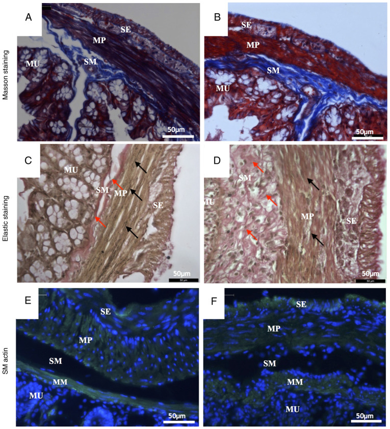
Ulcerative colitis (UC) is a complex disease that results from a dysregulated immune response in the gastrointestinal tract. A mouse model orally administered with dextran sodium sulfate (DSS) is the most widely used experimental animal model of UC. However, the ultrastructure of the colon in mouse colitis is poorly understood. In the present study, colonic specimens from DSS-induced UC mice underwent hematoxylin and eosin staining, Masson's trichrome staining and Verhoeff's elastic staining. In addition, the ultrastructure of samples was examined by transmission electron microscopy. UC was successfully induced by 7 consecutive days of DSS oral administration. The goblet cell architecture of the UC tissue was damaged in the mucosa. Additionally, a significant number of inflammatory cells infiltrated into the stroma and the structure of the intestinal gland was destroyed. The tissue in the submucosa showed significant edema. Hyperplasia was also identified in the submucosa, promoting a disorganized microstructure within the colon wall. Numerous collagen fibers in the muscular layer were disrupted, and the fiber bundles were thinner compared with those in the normal control group. Furthermore, in the DSS-induced UC group, the smooth muscle cell showed edema, the cell membrane structure was unclear and the shape of the nucleus was irregular. In conclusion, the present study revealed important histological and ultrastructural changes in the colon of DSS-induced UC mice. These features may contribute to improved understanding of the pathogenesis and mechanism of UC.
溃疡性结肠炎(UC)是一种复杂的疾病,由胃肠道免疫反应失调引起。口服葡聚糖硫酸钠(DSS)的小鼠模型是最广泛使用的UC实验动物模型。然而,小鼠结肠炎中结肠的超微结构尚不清楚。在本研究中,对DSS诱导的UC小鼠的结肠标本进行苏木精和伊红染色、Masson三色染色和Verhoeff弹性染色。此外,通过透射电子显微镜检查样品的超微结构。连续7天口服DSS成功诱导了UC。UC组织的杯状细胞结构在黏膜中受损。此外,大量炎性细胞浸润到基质中,肠腺结构被破坏。黏膜下层组织出现明显水肿。黏膜下层也发现增生,导致结肠壁内微观结构紊乱。肌层中大量胶原纤维被破坏,与正常对照组相比,纤维束更细。此外,在DSS诱导的UC组中,平滑肌细胞出现水肿,细胞膜结构不清晰,细胞核形状不规则。总之,本研究揭示了DSS诱导的UC小鼠结肠中重要的组织学和超微结构变化。这些特征可能有助于更好地理解UC的发病机制。