• 文献检索
  • 文档翻译
  • 深度研究
  • 学术资讯
  • Suppr Zotero 插件Zotero 插件
  • 邀请有礼
  • 套餐&价格
  • 历史记录
应用&插件
Suppr Zotero 插件Zotero 插件浏览器插件Mac 客户端Windows 客户端微信小程序
定价
高级版会员购买积分包购买API积分包
服务
文献检索文档翻译深度研究API 文档MCP 服务
关于我们
关于 Suppr公司介绍联系我们用户协议隐私条款
关注我们

Suppr 超能文献

核心技术专利:CN118964589B侵权必究
粤ICP备2023148730 号-1Suppr @ 2026

文献检索

告别复杂PubMed语法,用中文像聊天一样搜索,搜遍4000万医学文献。AI智能推荐,让科研检索更轻松。

立即免费搜索

文件翻译

保留排版,准确专业,支持PDF/Word/PPT等文件格式,支持 12+语言互译。

免费翻译文档

深度研究

AI帮你快速写综述,25分钟生成高质量综述,智能提取关键信息,辅助科研写作。

立即免费体验

巨噬细胞分泌的 MMP9 通过 PAR1 激活诱导胰腺癌细胞发生间质转化。

Macrophage-secreted MMP9 induces mesenchymal transition in pancreatic cancer cells via PAR1 activation.

机构信息

Center for Experimental and Molecular Medicine, Amsterdam UMC, University of Amsterdam, Amsterdam, The Netherlands.

Laboratory for Experimental Oncology and Radiobiology, Amsterdam UMC, University of Amsterdam, Cancer Center Amsterdam, Amsterdam, The Netherlands.

出版信息

Cell Oncol (Dordr). 2020 Dec;43(6):1161-1174. doi: 10.1007/s13402-020-00549-x. Epub 2020 Aug 18.

DOI:10.1007/s13402-020-00549-x
PMID:32809114
原文链接:https://pmc.ncbi.nlm.nih.gov/articles/PMC7717035/
Abstract

PURPOSE

Targeting tumor-infiltrating macrophages limits progression and improves chemotherapeutic responses in pancreatic ductal adenocarcinoma (PDAC). Protease-activated receptor (PAR)1 drives monocyte/macrophage recruitment, and stromal ablation of PAR1 limits cancer growth and enhances gemcitabine sensitivity in experimental PDAC. However, the functional interplay between PAR1, macrophages and tumor cells remains unexplored. Here we address the PAR1-macrophage-tumor cell crosstalk and assess its contributions to tumor progression.

METHODS

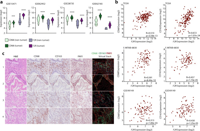
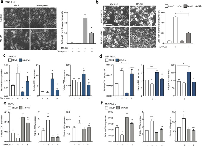
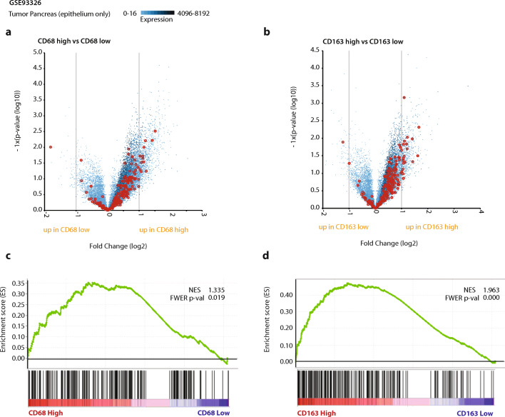
PAR1 expression and macrophage infiltration were correlated in primary PDAC biopsies using gene expression datasets and tissue microarrays. Medium transfer experiments were used to evaluate the functional consequences of macrophage-tumor cell crosstalk and to assess the contribution of PAR1 to the observed responses. PAR1 cleavage assays were used to identify a macrophage-secreted PAR1 agonist, and the effects of candidate proteases were assessed in medium transfer experiments with specific inhibitors and/or recombinant agonist.

RESULTS

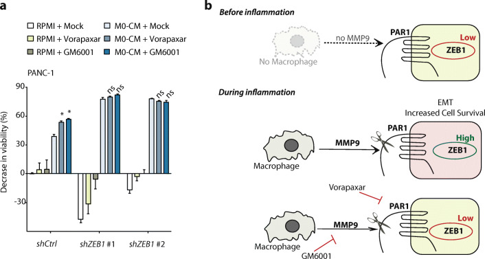
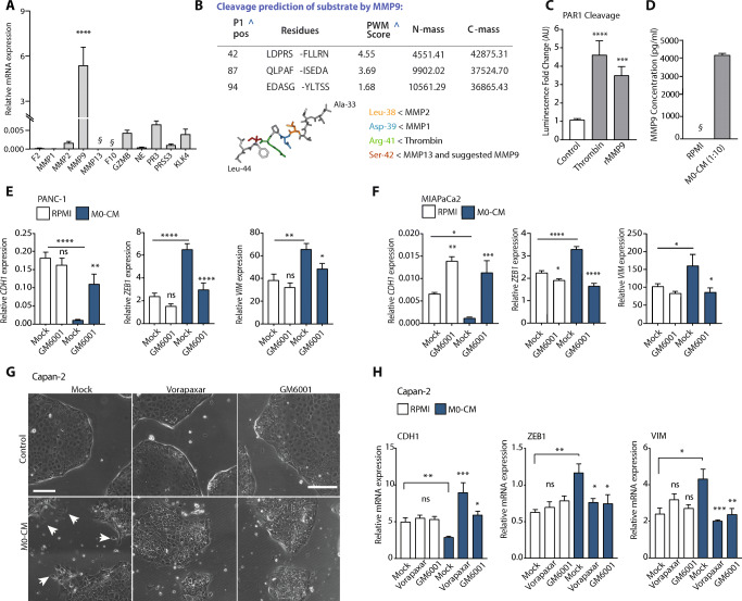
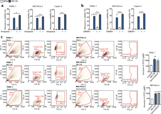
PAR1 expression correlates with macrophage infiltration in primary PDACs, and macrophages induce mesenchymal transition of PDAC cells through PAR1 activation. Protease profiling identified macrophage-secreted matrix metalloprotease 9 (MMP9) as the relevant PAR1 agonist in PDAC. PAR1 and/or MMP9 inhibition limited macrophage-driven mesenchymal transition. Likewise, preventing mesenchymal transition by silencing ZEB1 or by pharmacological inhibition of the MMP9/PAR1 axis significantly reduced the ability of tumor cells to survive the anti-tumor activities of macrophages.

CONCLUSION

Macrophages secrete MMP9, which acts upon PDAC cell PAR1 to induce mesenchymal transition. This macrophage-induced mesenchymal transition supports the tumor-promoting role of macrophage influx, explaining the dichotomous contributions of these immune cells to tumor growth.

摘要

目的

靶向肿瘤浸润巨噬细胞可限制胰腺导管腺癌(PDAC)的进展并提高化疗反应。蛋白酶激活受体(PAR)1 驱动单核细胞/巨噬细胞募集,基质中 PAR1 的消融可限制癌症生长并增强实验性 PDAC 中吉西他滨的敏感性。然而,PAR1、巨噬细胞和肿瘤细胞之间的功能相互作用仍未得到探索。在这里,我们研究了 PAR1-巨噬细胞-肿瘤细胞串扰,并评估了其对肿瘤进展的贡献。

方法

使用基因表达数据集和组织微阵列,在原发性 PDAC 活检中对 PAR1 表达和巨噬细胞浸润进行了相关性分析。采用介质转移实验来评估巨噬细胞-肿瘤细胞串扰的功能后果,并评估 PAR1 对观察到的反应的贡献。采用 PAR1 切割分析鉴定巨噬细胞分泌的 PAR1 激动剂,并在介质转移实验中使用特异性抑制剂和/或重组激动剂评估候选蛋白酶的作用。

结果

PAR1 表达与原发性 PDAC 中的巨噬细胞浸润相关,巨噬细胞通过 PAR1 激活诱导 PDAC 细胞发生间质转化。蛋白酶谱分析确定巨噬细胞分泌的基质金属蛋白酶 9(MMP9)是 PDAC 中相关的 PAR1 激动剂。PAR1 和/或 MMP9 抑制限制了巨噬细胞驱动的间质转化。同样,通过沉默 ZEB1 或通过 MMP9/PAR1 轴的药理学抑制来阻止间质转化,可显著降低肿瘤细胞对巨噬细胞抗肿瘤活性的生存能力。

结论

巨噬细胞分泌 MMP9,其作用于 PDAC 细胞 PAR1 以诱导间质转化。这种巨噬细胞诱导的间质转化支持巨噬细胞浸润的促肿瘤作用,解释了这些免疫细胞对肿瘤生长的双重贡献。

https://cdn.ncbi.nlm.nih.gov/pmc/blobs/18c8/7717035/f95c5316d285/13402_2020_549_Fig6_HTML.jpg
https://cdn.ncbi.nlm.nih.gov/pmc/blobs/18c8/7717035/f8b252b93ae9/13402_2020_549_Fig1_HTML.jpg
https://cdn.ncbi.nlm.nih.gov/pmc/blobs/18c8/7717035/14c8b8addc04/13402_2020_549_Fig2_HTML.jpg
https://cdn.ncbi.nlm.nih.gov/pmc/blobs/18c8/7717035/4ac7ae887fa1/13402_2020_549_Fig3_HTML.jpg
https://cdn.ncbi.nlm.nih.gov/pmc/blobs/18c8/7717035/b6294de6c7b2/13402_2020_549_Fig4_HTML.jpg
https://cdn.ncbi.nlm.nih.gov/pmc/blobs/18c8/7717035/293d2246ebc1/13402_2020_549_Fig5_HTML.jpg
https://cdn.ncbi.nlm.nih.gov/pmc/blobs/18c8/7717035/f95c5316d285/13402_2020_549_Fig6_HTML.jpg

相似文献

1
Macrophage-secreted MMP9 induces mesenchymal transition in pancreatic cancer cells via PAR1 activation.巨噬细胞分泌的 MMP9 通过 PAR1 激活诱导胰腺癌细胞发生间质转化。
Cell Oncol (Dordr). 2020 Dec;43(6):1161-1174. doi: 10.1007/s13402-020-00549-x. Epub 2020 Aug 18.
2
Protease-activated receptor 1 drives and maintains ductal cell fates in the premalignant pancreas and ductal adenocarcinoma.蛋白酶激活受体 1 驱动并维持癌前胰腺和导管腺癌中的导管细胞命运。
Mol Oncol. 2021 Nov;15(11):3091-3108. doi: 10.1002/1878-0261.12971. Epub 2021 May 14.
3
Endothelial-mesenchymal transition harnesses HSP90α-secreting M2-macrophages to exacerbate pancreatic ductal adenocarcinoma.内皮-间充质转化利用 HSP90α 分泌的 M2 巨噬细胞加剧胰腺导管腺癌。
J Hematol Oncol. 2019 Dec 17;12(1):138. doi: 10.1186/s13045-019-0826-2.
4
Thrombin-PAR1 signaling in pancreatic cancer promotes an immunosuppressive microenvironment.胰腺癌中的凝血酶-PAR1信号传导促进免疫抑制微环境。
J Thromb Haemost. 2021 Jan;19(1):161-172. doi: 10.1111/jth.15115. Epub 2020 Oct 25.
5
Tumor-driven like macrophages induced by conditioned media from pancreatic ductal adenocarcinoma promote tumor metastasis via secreting IL-8.肿瘤驱动型巨噬细胞由胰腺导管腺癌条件培养基诱导产生,通过分泌 IL-8 促进肿瘤转移。
Cancer Med. 2018 Nov;7(11):5679-5690. doi: 10.1002/cam4.1824. Epub 2018 Oct 12.
6
Knockdown of FOXO3a induces epithelial-mesenchymal transition and promotes metastasis of pancreatic ductal adenocarcinoma by activation of the β-catenin/TCF4 pathway through SPRY2.FOXO3a 的敲低通过 SPRY2 激活 β-catenin/TCF4 通路诱导胰腺导管腺癌的上皮-间充质转化并促进转移。
J Exp Clin Cancer Res. 2019 Jan 28;38(1):38. doi: 10.1186/s13046-019-1046-x.
7
PAR1 signaling on tumor cells limits tumor growth by maintaining a mesenchymal phenotype in pancreatic cancer.肿瘤细胞上的PAR1信号通过维持胰腺癌的间充质表型来限制肿瘤生长。
Oncotarget. 2018 Aug 10;9(62):32010-32023. doi: 10.18632/oncotarget.25880.
8
A Novel MIF Signaling Pathway Drives the Malignant Character of Pancreatic Cancer by Targeting NR3C2.一种新型的巨噬细胞移动抑制因子(MIF)信号通路通过靶向盐皮质激素受体(NR3C2)驱动胰腺癌的恶性特征。
Cancer Res. 2016 Jul 1;76(13):3838-50. doi: 10.1158/0008-5472.CAN-15-2841. Epub 2016 Apr 20.
9
Octyl Gallate Induces Pancreatic Ductal Adenocarcinoma Cell Apoptosis and Suppresses Endothelial-Mesenchymal Transition-Promoted M2-Macrophages, HSP90α Secretion, and Tumor Growth.辛基没食子酸酯诱导胰腺导管腺癌细胞凋亡,抑制内皮-间充质转化促进的 M2 巨噬细胞、HSP90α 分泌和肿瘤生长。
Cells. 2019 Dec 30;9(1):91. doi: 10.3390/cells9010091.
10
Interleukin 35 Expression Correlates With Microvessel Density in Pancreatic Ductal Adenocarcinoma, Recruits Monocytes, and Promotes Growth and Angiogenesis of Xenograft Tumors in Mice.白细胞介素 35 在胰腺导管腺癌中的表达与微血管密度相关,募集单核细胞,并促进小鼠异种移植肿瘤的生长和血管生成。
Gastroenterology. 2018 Feb;154(3):675-688. doi: 10.1053/j.gastro.2017.09.039. Epub 2017 Oct 6.

引用本文的文献

1
Domestication and feedback: bidirectional hijacking in pancreatic ductal adenocarcinoma microenvironment.驯化与反馈:胰腺导管腺癌微环境中的双向劫持
Front Immunol. 2025 Aug 11;16:1585858. doi: 10.3389/fimmu.2025.1585858. eCollection 2025.
2
Endogenous SLPI contributes to the regulation of inflammatory responses in peritoneal macrophages by modulating MMP-9 production.内源性分泌白细胞蛋白酶抑制因子通过调节基质金属蛋白酶-9的产生,有助于调节腹膜巨噬细胞中的炎症反应。
Front Immunol. 2025 May 27;16:1563845. doi: 10.3389/fimmu.2025.1563845. eCollection 2025.
3
Disulfidptosis classification of pancreatic carcinoma reveals correlation with clinical prognosis and immune profile.

本文引用的文献

1
Cancer statistics, 2019.癌症统计数据,2019 年。
CA Cancer J Clin. 2019 Jan;69(1):7-34. doi: 10.3322/caac.21551. Epub 2019 Jan 8.
2
PAR1 signaling on tumor cells limits tumor growth by maintaining a mesenchymal phenotype in pancreatic cancer.肿瘤细胞上的PAR1信号通过维持胰腺癌的间充质表型来限制肿瘤生长。
Oncotarget. 2018 Aug 10;9(62):32010-32023. doi: 10.18632/oncotarget.25880.
3
Protease-activated receptor-1 impedes prostate and intestinal tumor progression in mice.蛋白酶激活受体-1 抑制小鼠前列腺和肠道肿瘤的进展。
胰腺癌的二硫化物诱导细胞程序性坏死分类揭示了与临床预后和免疫特征的相关性。
Hereditas. 2025 Feb 22;162(1):26. doi: 10.1186/s41065-025-00381-z.
4
High levels of fibrotic tumor components are associated with recurrence and intratumoral immune status in advanced colorectal cancer patients.在晚期结直肠癌患者中,高水平的纤维化肿瘤成分与复发及肿瘤内免疫状态相关。
Sci Rep. 2024 Dec 28;14(1):30735. doi: 10.1038/s41598-024-80489-w.
5
Single-nuclei transcriptome analysis of channel catfish spleen provides insight into the immunome of an aquaculture-relevant species.对斑点叉尾鮰脾脏的单细胞转录组分析为水产养殖相关物种的免疫组提供了新见解。
PLoS One. 2024 Sep 26;19(9):e0309397. doi: 10.1371/journal.pone.0309397. eCollection 2024.
6
Recombinant hirudin and PAR-1 regulate macrophage polarisation status in diffuse large B-cell lymphoma.重组水蛭素和 PAR-1 调节弥漫性大 B 细胞淋巴瘤中巨噬细胞的极化状态。
BMC Biotechnol. 2024 Aug 12;24(1):55. doi: 10.1186/s12896-024-00879-w.
7
Macrophage heterogeneity and its interactions with stromal cells in tumour microenvironment.肿瘤微环境中巨噬细胞的异质性及其与基质细胞的相互作用。
Cell Biosci. 2024 Feb 1;14(1):16. doi: 10.1186/s13578-024-01201-z.
8
Differential Effects of Pancreatic Cancer-Derived Extracellular Vesicles Driving a Suppressive Environment.胰腺癌衍生细胞外囊泡诱导抑制微环境的差异效应。
Int J Mol Sci. 2023 Sep 27;24(19):14652. doi: 10.3390/ijms241914652.
9
Comparative analysis of single-cell transcriptome reveals heterogeneity in the tumor microenvironment of lung adenocarcinoma and brain metastases.单细胞转录组的比较分析揭示了肺腺癌和脑转移瘤微环境中的异质性。
Discov Oncol. 2023 Sep 15;14(1):174. doi: 10.1007/s12672-023-00784-2.
10
Integration of Single-Cell RNA Sequencing and Bulk RNA Sequencing Reveals That TAM2-Driven Genes Affect Immunotherapeutic Response and Prognosis in Pancreatic Cancer.单细胞 RNA 测序和批量 RNA 测序的整合表明,TAM2 驱动的基因影响胰腺癌的免疫治疗反应和预后。
Int J Mol Sci. 2023 Aug 14;24(16):12787. doi: 10.3390/ijms241612787.
J Thromb Haemost. 2018 Nov;16(11):2258-2269. doi: 10.1111/jth.14277. Epub 2018 Sep 27.
4
Andecaliximab/GS-5745 Alone and Combined with mFOLFOX6 in Advanced Gastric and Gastroesophageal Junction Adenocarcinoma: Results from a Phase I Study.安可达西单抗/GS-5745 单药及联合 mFOLFOX6 治疗晚期胃及胃食管结合部腺癌的 I 期研究结果。
Clin Cancer Res. 2018 Aug 15;24(16):3829-3837. doi: 10.1158/1078-0432.CCR-17-2469. Epub 2018 Apr 24.
5
Neutrophil Extracellular Traps Participate in All Different Types of Thrombotic and Haemorrhagic Complications of Coronary Atherosclerosis.中性粒细胞胞外陷阱参与冠状动脉粥样硬化的所有不同类型的血栓和出血并发症。
Thromb Haemost. 2018 Jun;118(6):1078-1087. doi: 10.1055/s-0038-1641749. Epub 2018 Apr 19.
6
Comprehensive characterisation of compartment-specific long non-coding RNAs associated with pancreatic ductal adenocarcinoma.全面描绘与胰腺导管腺癌相关的特定隔室长非编码 RNA。
Gut. 2019 Mar;68(3):499-511. doi: 10.1136/gutjnl-2017-314353. Epub 2018 Feb 10.
7
Targeting PAR1: Now What?靶向PAR1:接下来怎么办?
Trends Pharmacol Sci. 2017 Aug;38(8):701-716. doi: 10.1016/j.tips.2017.05.001. Epub 2017 May 27.
8
Metalloproteinase-9 contributes to endothelial dysfunction in atherosclerosis via protease activated receptor-1.金属蛋白酶-9通过蛋白酶激活受体-1导致动脉粥样硬化中的内皮功能障碍。
PLoS One. 2017 Feb 6;12(2):e0171427. doi: 10.1371/journal.pone.0171427. eCollection 2017.
9
Endothelial Nitric Oxide Synthase Traffic Inducer (NOSTRIN) is a Negative Regulator of Disease Aggressiveness in Pancreatic Cancer.内皮型一氧化氮合酶转运诱导因子(NOSTRIN)是胰腺癌疾病侵袭性的负调节因子。
Clin Cancer Res. 2016 Dec 15;22(24):5992-6001. doi: 10.1158/1078-0432.CCR-16-0511. Epub 2016 Jul 8.
10
Macrophages and pancreatic ductal adenocarcinoma.巨噬细胞与胰腺导管腺癌
Cancer Lett. 2016 Oct 10;381(1):211-6. doi: 10.1016/j.canlet.2015.11.049. Epub 2015 Dec 18.