Department of Chemical and Biological Physics, Weizmann Institute, Rehovot, Israel.
Division of Physical Chemistry, Department of Chemistry, Lund University, Lund, Sweden.
NMR Biomed. 2020 Nov;33(11):e4355. doi: 10.1002/nbm.4355. Epub 2020 Aug 19.
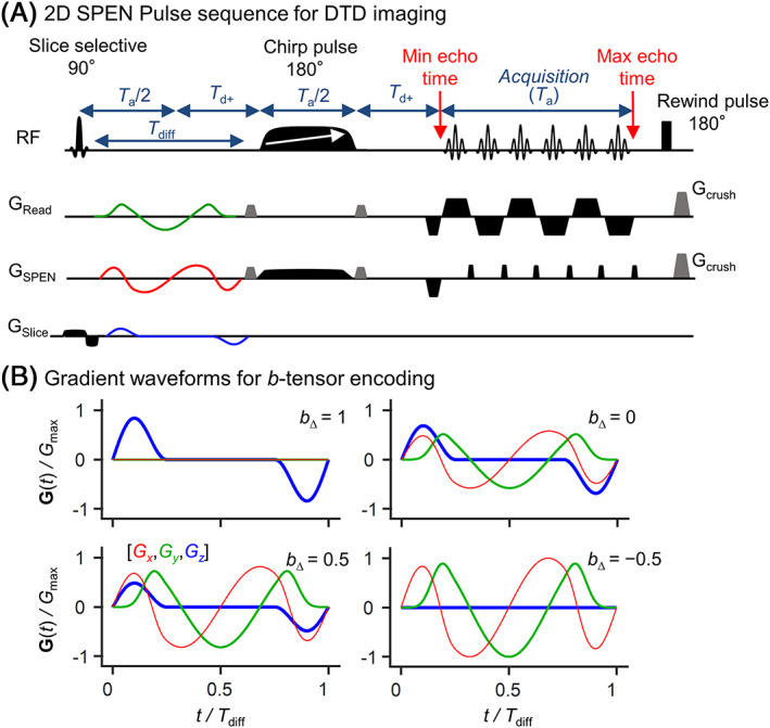
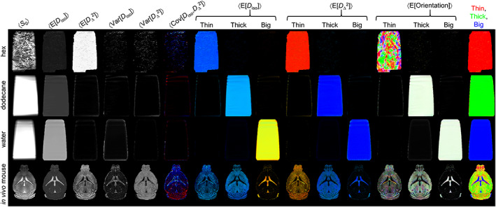
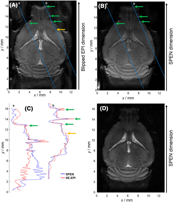
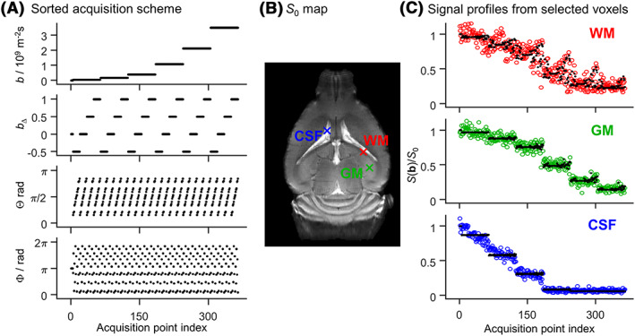
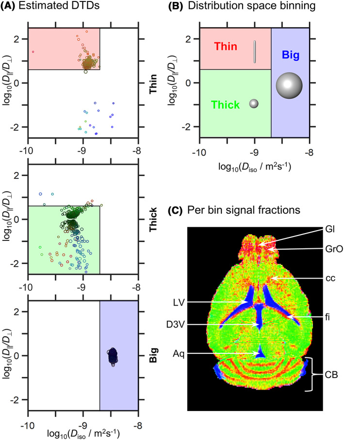
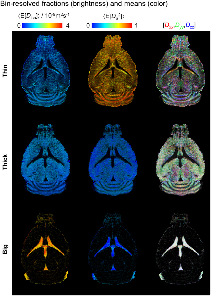
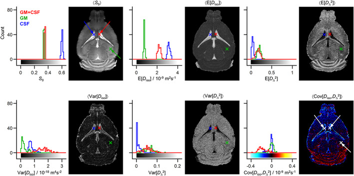
Diffusion tensor distribution (DTD) imaging builds on principles from diffusion, solid-state and low-field NMR spectroscopies, to quantify the contents of heterogeneous voxels as nonparametric distributions, with tensor "size", "shape" and orientation having direct relations to corresponding microstructural properties of biological tissues. The approach requires the acquisition of multiple images as a function of the magnitude, shape and direction of the diffusion-encoding gradients, leading to long acquisition times unless fast image read-out techniques like EPI are employed. While in previous in vivo human brain studies performed at 3 T this proved a viable option, porting these measurements to very high magnetic fields and/or to heterogeneous organs induces B - and B -inhomogeneity artifacts that challenge the limits of EPI. To overcome such challenges, we demonstrate here that high spatial resolution DTD of mouse brain can be carried out at 15.2 T with a surface-cryoprobe, by relying on SPatiotemporal ENcoding (SPEN) imaging sequences. These new acquisition and data-processing protocols are demonstrated with measurements on in vivo mouse brain, and validated with synthetic phantoms designed to mimic the diffusion properties of white matter, gray matter and cerebrospinal fluid. While still in need of full extensions to 3D mappings and of scanning additional animals to extract more general physiological conclusions, this work represents another step towards the model-free, noninvasive in vivo characterization of tissue microstructure and heterogeneity in animal models, at ≈0.1 mm resolutions.
弥散张量分布(DTD)成像基于扩散、固态和低场 NMR 光谱学原理,对不均匀体素的内容进行量化,作为非参数分布,张量“大小”、“形状”和方向与生物组织的相应微观结构特性直接相关。该方法需要获取多个图像,作为扩散编码梯度的大小、形状和方向的函数,导致采集时间长,除非使用快速图像读出技术如 EPI。虽然在之前在 3T 进行的人体大脑研究中,这是一种可行的选择,但将这些测量方法移植到非常高的磁场和/或不均匀的器官中,会引起 B -和 B -不均匀性伪影,这对 EPI 的极限提出了挑战。为了克服这些挑战,我们在此展示了通过依赖于时空编码(SPEN)成像序列,可以在 15.2T 用表面冷冻探头对小鼠大脑进行高空间分辨率的 DTD。这些新的采集和数据处理协议通过对体内小鼠大脑的测量进行了演示,并通过设计来模拟白质、灰质和脑脊液的扩散特性的合成体模进行了验证。虽然仍需要全面扩展到 3D 映射,并扫描更多的动物以提取更普遍的生理结论,但这项工作代表了朝着在动物模型中进行无模型、非侵入性的组织微观结构和异质性的体内特征化的又一步,分辨率约为 0.1mm。