Tissue Engineering and Stem Cell Group, Singapore Eye Research Institute, Singapore.
Singapore National Eye Centre, Singapore.
Transl Vis Sci Technol. 2020 Mar 9;9(4):6. doi: 10.1167/tvst.9.4.6. eCollection 2020 Mar.
To investigate the postoperative inflammatory and wound-healing responses after laser scleral microporation for presbyopia.
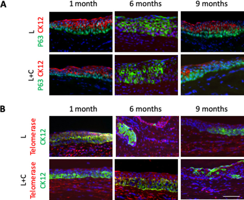
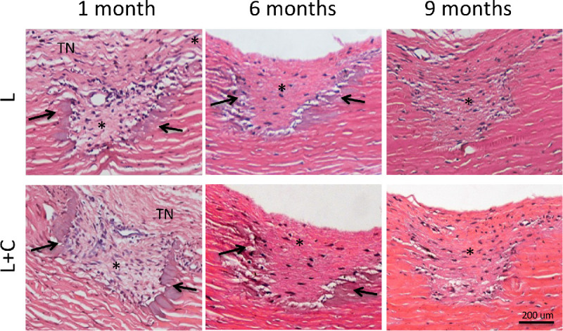
Thirty porcine eyes were used for the optimization of laser intensities first. Six monkeys (12 eyes) received scleral microporation with an erbium yttrium aluminum garnet (Er:YAG) laser, and half of the eyes received concurrent subconjunctival collagen gel to modulate wound-healing response. The intraocular pressure (IOP) and the laser ablation depth were evaluated. The animals were euthanized at 1, 6, and 9 months postoperatively. The limbal areas and scleras were harvested for histologic analysis and immunofluorescence of markers for inflammation (CD11b and CD45), wound healing (CD90, tenascin-C, fibronectin, and HSP47), wound contraction (α-smooth muscle actin [α-SMA]), vascular response (CD31), nerve injury (GAP43), and limbal stem cells (P63 and telomerase).
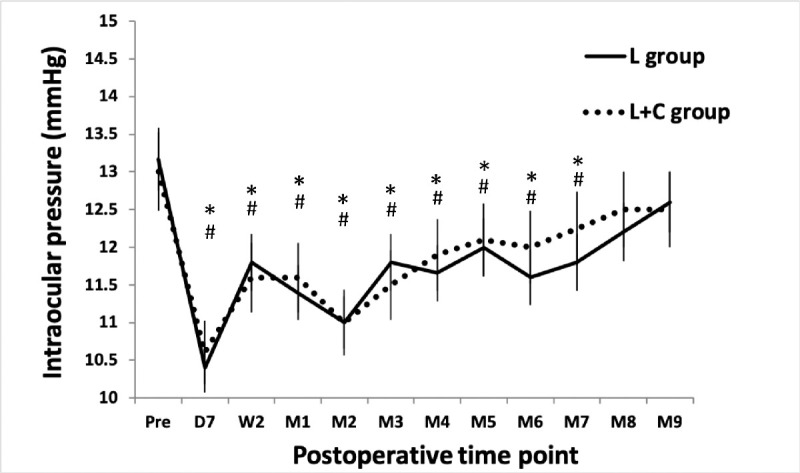
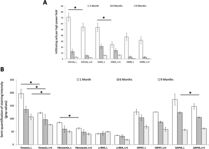
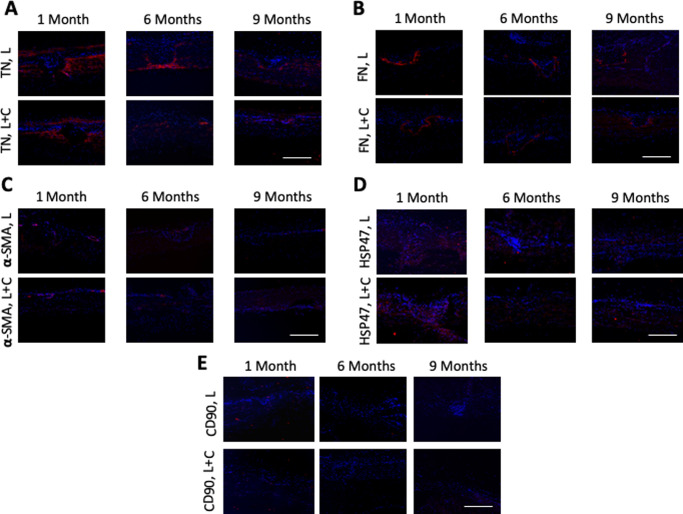
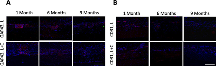
In the nonhuman primate study, there was a significant reduction in IOP after the procedure. Overall, the ablation depth was 76.6% to 81.2% at 1 month and slightly decreased to 71.5% to 72.7% at 9 months. Coagulative necrosis around the micropores, as well as expression of CD11b, CD45, tenascin, fibronectin, HSP47, and GAP43, was distinct at 1 month but subsided with time. Collagen gel treatment significantly suppressed the upregulation of CD11b, CD45, fibronectin, and tenascin-C. The expression of CD90, α-SMA, and CD31 was minimal in all eyes.
The study demonstrated the course of inflammatory and wound-healing responses following laser scleral microporation. The tissue responses were small and self-limited, resolved with time, and were suppressed by concurrent collagen treatment. It provides a useful understanding of this new procedure.
The results would be helpful in the laser parameter modification to improve the long-term treatment stability.
研究激光巩膜微孔成形术治疗老视术后的炎症和伤口愈合反应。
首先用 30 只猪眼优化激光强度。6 只猴子(12 只眼)接受铒石榴石激光(Er:YAG)巩膜微孔成形术,其中一半眼接受结膜下胶原凝胶以调节伤口愈合反应。评估眼内压(IOP)和激光消融深度。术后 1、6 和 9 个月处死动物。采集角膜缘区和巩膜进行组织学分析和炎症标志物(CD11b 和 CD45)、伤口愈合(CD90、腱糖蛋白-C、纤维连接蛋白和 HSP47)、伤口收缩(α-平滑肌肌动蛋白[α-SMA])、血管反应(CD31)、神经损伤(GAP43)和角膜缘干细胞(P63 和端粒酶)的免疫荧光。
在非人类灵长类动物研究中,术后 IOP 显著降低。总体而言,1 个月时的消融深度为 76.6%至 81.2%,9 个月时略有下降至 71.5%至 72.7%。微孔周围的凝固性坏死以及 CD11b、CD45、腱糖蛋白、纤维连接蛋白、HSP47 和 GAP43 的表达在 1 个月时明显,但随时间消退。胶原凝胶治疗显著抑制了 CD11b、CD45、纤维连接蛋白和腱糖蛋白-C 的上调。所有眼中 CD90、α-SMA 和 CD31 的表达均较少。
该研究表明了激光巩膜微孔成形术后炎症和伤口愈合反应的过程。组织反应较小且自限性,随时间消退,并通过同时进行胶原治疗得到抑制。它为这种新手术提供了有用的理解。
严佳鑫