• 文献检索
  • 文档翻译
  • 深度研究
  • 学术资讯
  • Suppr Zotero 插件Zotero 插件
  • 邀请有礼
  • 套餐&价格
  • 历史记录
应用&插件
Suppr Zotero 插件Zotero 插件浏览器插件Mac 客户端Windows 客户端微信小程序
定价
高级版会员购买积分包购买API积分包
服务
文献检索文档翻译深度研究API 文档MCP 服务
关于我们
关于 Suppr公司介绍联系我们用户协议隐私条款
关注我们

Suppr 超能文献

核心技术专利:CN118964589B侵权必究
粤ICP备2023148730 号-1Suppr @ 2026

文献检索

告别复杂PubMed语法,用中文像聊天一样搜索,搜遍4000万医学文献。AI智能推荐,让科研检索更轻松。

立即免费搜索

文件翻译

保留排版,准确专业,支持PDF/Word/PPT等文件格式,支持 12+语言互译。

免费翻译文档

深度研究

AI帮你快速写综述,25分钟生成高质量综述,智能提取关键信息,辅助科研写作。

立即免费体验

不同深度距骨骨软骨损伤对踝关节稳定性影响的有限元分析

Finite Element Analysis of the Effect of Talar Osteochondral Defects of Different Depths on Ankle Joint Stability.

作者信息

Li Jia, Wei Yu, Wei Min

机构信息

Department of Orthopedics, Chinese People's Liberation Army (PLA) General Hospital, Beijing, China (mainland).

出版信息

Med Sci Monit. 2020 Aug 21;26:e921823. doi: 10.12659/MSM.921823.

DOI:10.12659/MSM.921823
PMID:32820745
原文链接:https://pmc.ncbi.nlm.nih.gov/articles/PMC7456163/
Abstract

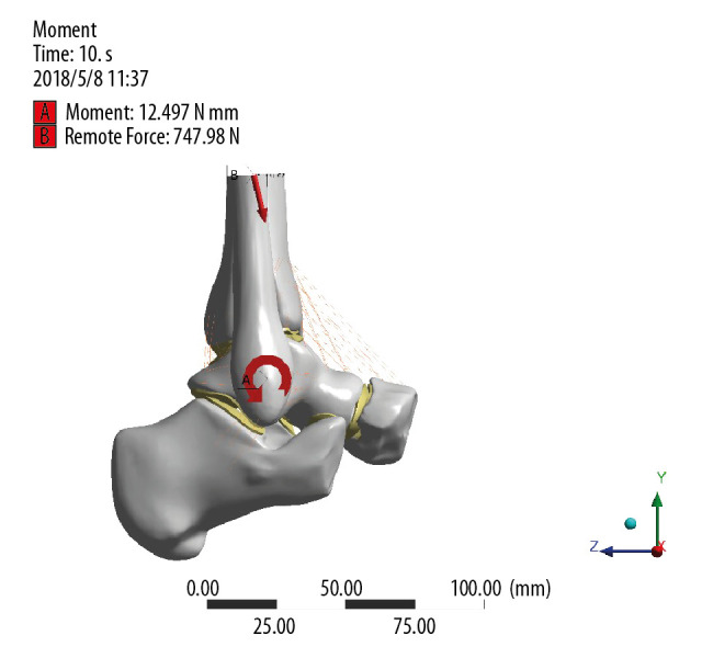
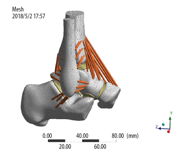
BACKGROUND Talus cartilage injury leads to changes in biomechanics of the ankle joint and ultimately affects ankle joint function, but which talus cartilage defects require surgery is still uncertain. This research used a finite element method to simulate the effect of different depth of talus cartilage defects on the stress and stability of the ankle joint in a certain area. MATERIAL AND METHODS A three-dimensional finite element model with different depths of osteochondral defects was created to simulate and calculate joint stress and displacement of the articular surface of the distal tibia and the proximal talus while the ankle joint was in the push-off, midstance, and heel-strike phases. RESULTS The equivalent stress of the proximal talus did not change significantly at a defect depth of 1 mm, whereas the equivalent stress of the upper talus increased significantly at a defect depth of ≥3 mm or more, reaching a maximum value at a defect depth of 10 mm. The equivalent stress of the tibial cartilage and the equivalent stress and displacement in the corresponding forces in the midstance phase and heel-strike phase were significantly different from those in the normal group, but the difference in stress in each defect group was not obvious. CONCLUSIONS The effect of cartilage defects of the talus on biomechanics of the ankle is clear, especially in the midstance and push-off phases. When the defect reaches the subchondral bone (at a depth of 3 mm), the most obvious change in ankle joint stability occurs, and it does not increase linearly with the increase in depth of the defect.

摘要

背景 距骨软骨损伤会导致踝关节生物力学改变,最终影响踝关节功能,但哪些距骨软骨缺损需要手术仍不明确。本研究采用有限元方法模拟距骨软骨缺损不同深度对踝关节特定区域应力和稳定性的影响。

材料与方法 建立具有不同深度骨软骨缺损的三维有限元模型,以模拟和计算踝关节在蹬离期、站立中期和足跟撞击期时胫骨远端和距骨近端关节面的关节应力和位移。

结果 缺损深度为1mm时,距骨近端的等效应力无明显变化,而缺损深度≥3mm及以上时,距骨上部的等效应力显著增加,在缺损深度为10mm时达到最大值。胫骨软骨的等效应力以及站立中期和足跟撞击期相应力中的等效应力和位移与正常组有显著差异,但各缺损组间应力差异不明显。

结论 距骨软骨缺损对踝关节生物力学的影响明显,尤其是在站立中期和蹬离期。当缺损累及软骨下骨(深度为3mm)时,踝关节稳定性变化最为明显,且并不随缺损深度增加呈线性增加。

https://cdn.ncbi.nlm.nih.gov/pmc/blobs/b3ed/7456163/6c97bd52527a/medscimonit-26-e921823-g004.jpg
https://cdn.ncbi.nlm.nih.gov/pmc/blobs/b3ed/7456163/4e1f7b7f1a90/medscimonit-26-e921823-g001.jpg
https://cdn.ncbi.nlm.nih.gov/pmc/blobs/b3ed/7456163/1ce0e3b2c8a4/medscimonit-26-e921823-g002.jpg
https://cdn.ncbi.nlm.nih.gov/pmc/blobs/b3ed/7456163/b6b8dbd2e4d2/medscimonit-26-e921823-g003.jpg
https://cdn.ncbi.nlm.nih.gov/pmc/blobs/b3ed/7456163/6c97bd52527a/medscimonit-26-e921823-g004.jpg

相似文献

1
Finite Element Analysis of the Effect of Talar Osteochondral Defects of Different Depths on Ankle Joint Stability.不同深度距骨骨软骨损伤对踝关节稳定性影响的有限元分析
Med Sci Monit. 2020 Aug 21;26:e921823. doi: 10.12659/MSM.921823.
2
The effect of talus osteochondral defects of different area size on ankle joint stability: a finite element analysis.不同面积大小距骨骨软骨缺损对踝关节稳定性的影响:有限元分析。
BMC Musculoskelet Disord. 2022 May 27;23(1):500. doi: 10.1186/s12891-022-05450-2.
3
The Biomechanical Influence of Defected Cartilage on the Progression of Osteochondral Lesions of the Talus: A Three-dimensional Finite Element Analysis.软骨缺损对距骨骨软骨病变进展的生物力学影响:三维有限元分析。
Orthop Surg. 2023 Jun;15(6):1685-1693. doi: 10.1111/os.13753. Epub 2023 May 17.
4
The biomechanical influence of tibio-talar containment on stability of the ankle joint.胫距包容对踝关节稳定性的生物力学影响。
Knee Surg Sports Traumatol Arthrosc. 2007 Nov;15(11):1355-62. doi: 10.1007/s00167-007-0372-2. Epub 2007 Jul 13.
5
Histomorphometric, CT arthrographic, and biomechanical mapping of the human ankle.人体踝关节的组织形态计量学、CT 关节造影和生物力学图谱。
Foot Ankle Int. 2013 Jul;34(7):1025-34. doi: 10.1177/1071100713477636. Epub 2013 Feb 8.
6
Evaluation of Articular Cartilage Injury Using Computed Tomography With Axial Traction in the Ankle Joint.使用踝关节轴向牵引计算机断层扫描评估关节软骨损伤
Foot Ankle Int. 2018 Sep;39(9):1120-1127. doi: 10.1177/1071100718777489. Epub 2018 May 18.
7
Treatment of talus osteochondral defects in chronic lateral unstable ankles: small-sized lateral chondral lesions had good clinical outcomes.慢性外不稳定踝关节距骨骨软骨缺损的治疗:小面积外侧软骨病变有良好的临床疗效。
Knee Surg Sports Traumatol Arthrosc. 2018 Jul;26(7):2116-2122. doi: 10.1007/s00167-017-4591-x. Epub 2017 Jun 2.
8
The Influence of Talar Displacement on Articular Contact Mechanics: A 3D Finite Element Analysis Study Using Weightbearing Computed Tomography.距骨位移对关节接触力学的影响:基于负重位 CT 的三维有限元分析研究。
Foot Ankle Int. 2024 Apr;45(4):393-405. doi: 10.1177/10711007241227179. Epub 2024 Feb 25.
9
Effect of cartilage thickness mismatch in osteochondral grafting from knee to talus on articular contact pressures: A finite element analysis.膝关节至距骨骨软骨移植中软骨厚度不匹配对关节接触压力的影响:有限元分析
Jt Dis Relat Surg. 2021;32(2):355-362. doi: 10.52312/jdrs.2021.41. Epub 2021 Jun 11.
10
Cartilage thickness in cadaveric ankles: measurement with double-contrast multi-detector row CT arthrography versus MR imaging.尸体踝关节软骨厚度:双对比多层螺旋CT关节造影与磁共振成像测量对比
Radiology. 2004 Dec;233(3):768-73. doi: 10.1148/radiol.2333031921. Epub 2004 Oct 29.

引用本文的文献

1
Osteochondral lesions of talus.距骨骨软骨损伤
Bone Joint Res. 2024 Dec 17;13(12):790-792. doi: 10.1302/2046-3758.1312.BJR-2024-0503.
2
Visualized trends and bibliometric analysis in ankle cartilage repair from 2004 to 2024.2004年至2024年踝关节软骨修复的可视化趋势与文献计量分析
Front Med (Lausanne). 2024 Nov 20;11:1503707. doi: 10.3389/fmed.2024.1503707. eCollection 2024.
3
Investigation into the effect of deltoid ligament injury on rotational ankle instability using a three-dimensional ankle finite element model.

本文引用的文献

1
Arthroscopic Microfracture for Osteochondral Lesions of the Talus: Functional Outcomes at a Mean of 6.7 Years in 165 Consecutive Ankles.关节镜下微骨折术治疗距骨骨软骨病变:165 例连续踝关节平均 6.7 年的功能结果。
Am J Sports Med. 2020 Jan;48(1):153-158. doi: 10.1177/0363546519887957.
2
Midterm Outcomes of Bone Marrow Stimulation for Primary Osteochondral Lesions of the Talus: A Systematic Review.距骨原发性骨软骨损伤的骨髓刺激中期结果:一项系统评价
Orthop J Sports Med. 2019 Oct 30;7(10):2325967119879127. doi: 10.1177/2325967119879127. eCollection 2019 Oct.
3
The use of wearable devices for walking and running gait analysis outside of the lab: A systematic review.
使用三维踝关节有限元模型研究三角韧带损伤对踝关节旋转不稳的影响。
Front Bioeng Biotechnol. 2024 May 1;12:1386401. doi: 10.3389/fbioe.2024.1386401. eCollection 2024.
4
The Biomechanical Influence of Defected Cartilage on the Progression of Osteochondral Lesions of the Talus: A Three-dimensional Finite Element Analysis.软骨缺损对距骨骨软骨病变进展的生物力学影响:三维有限元分析。
Orthop Surg. 2023 Jun;15(6):1685-1693. doi: 10.1111/os.13753. Epub 2023 May 17.
5
Osteochondritis dissecans of the metacarpal head in a soldier treated with osteochondral autograft transplantation surgery: A case report.掌骨头骨软骨病损士兵采用骨软骨自体移植手术治疗:1 例报告。
Medicine (Baltimore). 2023 Jan 20;102(3):e32563. doi: 10.1097/MD.0000000000032563.
6
The effect of talus osteochondral defects of different area size on ankle joint stability: a finite element analysis.不同面积大小距骨骨软骨缺损对踝关节稳定性的影响:有限元分析。
BMC Musculoskelet Disord. 2022 May 27;23(1):500. doi: 10.1186/s12891-022-05450-2.
7
Effect of cartilage thickness mismatch in osteochondral grafting from knee to talus on articular contact pressures: A finite element analysis.膝关节至距骨骨软骨移植中软骨厚度不匹配对关节接触压力的影响:有限元分析
Jt Dis Relat Surg. 2021;32(2):355-362. doi: 10.52312/jdrs.2021.41. Epub 2021 Jun 11.
实验室外可穿戴设备在步行和跑步步态分析中的应用:一项系统综述。
Gait Posture. 2018 Jun;63:124-138. doi: 10.1016/j.gaitpost.2018.04.047. Epub 2018 May 1.
4
Proteomic analysis of synovial fluid in osteoarthritis using SWATH‑mass spectrometry.采用 SWATH 质谱法对骨关节炎滑液进行蛋白质组学分析。
Mol Med Rep. 2018 Feb;17(2):2827-2836. doi: 10.3892/mmr.2017.8250. Epub 2017 Dec 11.
5
Management of Osteochondral Lesions of the Talar Dome.距骨穹窿骨软骨损伤的管理
Open Orthop J. 2017 Jul 31;11:743-761. doi: 10.2174/1874325001711010743. eCollection 2017.
6
Osteochondral lesions of the talus in the athlete: up to date review.运动员距骨的骨软骨损伤:最新综述
Curr Rev Musculoskelet Med. 2017 Mar;10(1):131-140. doi: 10.1007/s12178-017-9393-8.
7
Current management of talar osteochondral lesions.距骨骨软骨损伤的当前治疗方法。
World J Orthop. 2017 Jan 18;8(1):12-20. doi: 10.5312/wjo.v8.i1.12.
8
Osteochondral autograft transplantation with biplanar distal tibial osteotomy for patients with concomitant large osteochondral lesion of the talus and varus ankle malalignment.采用双平面胫骨远端截骨术进行自体骨软骨移植治疗合并距骨大骨软骨损伤和内翻踝关节畸形的患者。
BMC Musculoskelet Disord. 2017 Jan 19;18(1):23. doi: 10.1186/s12891-016-1367-2.
9
Evaluation and Management of Osteochondral Lesions of the Talus.距骨骨软骨损伤的评估与处理
Cartilage. 2017 Jan;8(1):19-30. doi: 10.1177/1947603516670708. Epub 2016 Sep 28.
10
Lesion Size Is a Predictor of Clinical Outcomes After Bone Marrow Stimulation for Osteochondral Lesions of the Talus: A Systematic Review.病变大小是距骨骨软骨损伤骨髓刺激术后临床结果的预测指标:一项系统评价。
Am J Sports Med. 2017 Jun;45(7):1698-1705. doi: 10.1177/0363546516668292. Epub 2016 Nov 16.