Division of Cancer Immunology, Research Institute, National Cancer Center, Tokyo, Japan.
Exploratory Oncology Research and Clinical Trial Center (EPOC), National Cancer Center, Kashiwa, Japan.
Blood Adv. 2020 Sep 8;4(17):4069-4082. doi: 10.1182/bloodadvances.2020002098.
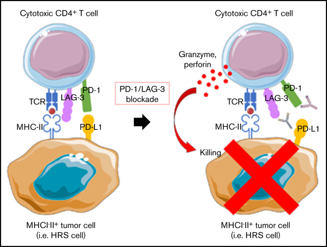
Classic Hodgkin lymphoma (cHL) responds markedly to PD-1 blockade therapy, and the clinical responses are reportedly dependent on expression of major histocompatibility complex class II (MHC-II). This dependence is different from other solid tumors, in which the MHC class I (MHC-I)/CD8+ T-cell axis plays a critical role. In this study, we investigated the role of the MHC-II/CD4+ T-cell axis in the antitumor effect of PD-1 blockade on cHL. In cHL, MHC-I expression was frequently lost, but MHC-II expression was maintained. CD4+ T cells highly infiltrated the tumor microenvironment of MHC-II-expressing cHL, regardless of MHC-I expression status. Consequently, CD4+ T-cell, but not CD8+ T-cell, infiltration was a good prognostic factor in cHL, and PD-1 blockade showed antitumor efficacy against MHC-II-expressing cHL associated with CD4+ T-cell infiltration. Murine lymphoma and solid tumor models revealed the critical role of antitumor effects mediated by CD4+ T cells: an anti-PD-1 monoclonal antibody exerted antitumor effects on MHC-I-MHC-II+ tumors but not on MHC-I-MHC-II- tumors, in a cytotoxic CD4+ T-cell-dependent manner. Furthermore, LAG-3, which reportedly binds to MHC-II, was highly expressed by tumor-infiltrating CD4+ T cells in MHC-II-expressing tumors. Therefore, the combination of LAG-3 blockade with PD-1 blockade showed a far stronger antitumor immunity compared with either treatment alone. We propose that PD-1 blockade therapies have antitumor effects on MHC-II-expressing tumors such as cHL that are mediated by cytotoxic CD4+ T cells and that LAG-3 could be a candidate for combination therapy with PD-1 blockade.
经典型霍奇金淋巴瘤 (cHL) 对 PD-1 阻断治疗有明显反应,据报道,临床反应取决于主要组织相容性复合体 II (MHC-II) 的表达。这种依赖性与其他实体瘤不同,在实体瘤中,MHC 类 I (MHC-I)/CD8+ T 细胞轴起着关键作用。在这项研究中,我们研究了 MHC-II/CD4+ T 细胞轴在 PD-1 阻断对 cHL 的抗肿瘤作用中的作用。在 cHL 中,MHC-I 的表达经常丢失,但 MHC-II 的表达得到维持。CD4+ T 细胞高度浸润 MHC-II 表达的 cHL 肿瘤微环境,无论 MHC-I 的表达状态如何。因此,CD4+ T 细胞浸润而非 CD8+ T 细胞浸润是 cHL 的良好预后因素,PD-1 阻断对与 CD4+ T 细胞浸润相关的 MHC-II 表达的 cHL 显示出抗肿瘤疗效。鼠淋巴瘤和实体瘤模型揭示了 CD4+ T 细胞介导的抗肿瘤作用的关键作用:抗 PD-1 单克隆抗体对 MHC-I-MHC-II+ 肿瘤发挥抗肿瘤作用,但对 MHC-I-MHC-II- 肿瘤没有作用,这是一种依赖于细胞毒性 CD4+ T 细胞的方式。此外,LAG-3 据报道与 MHC-II 结合,在 MHC-II 表达的肿瘤中高度表达于肿瘤浸润的 CD4+ T 细胞。因此,与单独治疗相比,LAG-3 阻断与 PD-1 阻断的联合治疗显示出更强的抗肿瘤免疫作用。我们提出,PD-1 阻断疗法对 MHC-II 表达的肿瘤(如 cHL)具有抗肿瘤作用,这种作用是由细胞毒性 CD4+ T 细胞介导的,LAG-3 可能是与 PD-1 阻断联合治疗的候选药物。