Molecular Mechanisms of Brain Development, Center for Brain Science (CBS), RIKEN, Saitama, Japan.
Exploratory Research for Advanced Technology (ERATO), Japan Science and Technology Agency (JST), Tokyo, Japan.
Sci Rep. 2020 Sep 2;10(1):14437. doi: 10.1038/s41598-020-71474-0.
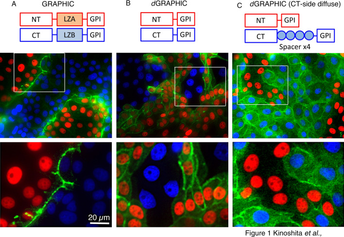
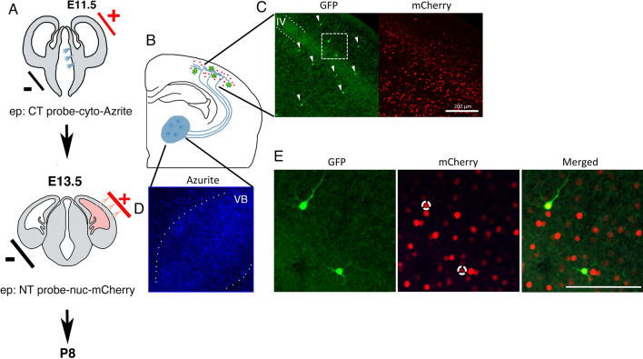
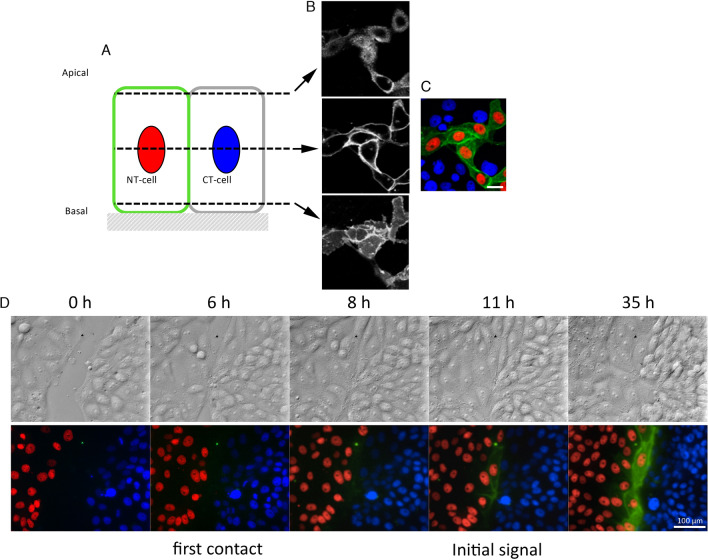
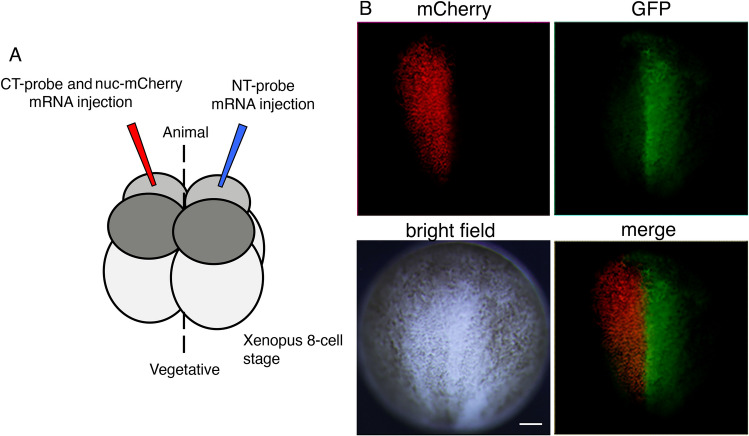
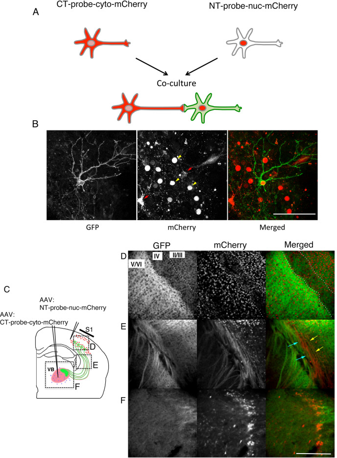
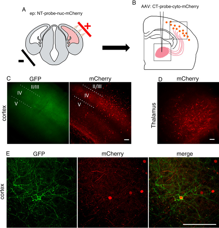
The ability to identify specific cell-cell contact in the highly heterogeneous mammalian body is crucial to revealing precise control of the body plan and correct function. To visualize local connections, we previously developed a genetically encoded fluorescent indicator, GRAPHIC, which labels cell-cell contacts by restricting the reconstituted green fluorescent protein (GFP) signal to the contact site. Here, we modify GRAPHIC to give the reconstituted GFP motility within the membrane, to detect cells that make contact with other specific cells. Removal of leucine zipper domains, located between the split GFP fragment and glycophosphatidylinositol anchor domain, allowed GFP reconstituted at the contact site to diffuse throughout the entire plasma membrane, revealing cell morphology. Further, depending on the structural spacers employed, the reconstituted GFP could be selectively targeted to N terminal (NT)- or C terminal (CT)-probe-expressing cells. Using these novel constructs, we demonstrated that we can specifically label NT-probe-expressing cells that made contact with CT-probe-expressing cells in an epithelial cell culture and in Xenopus 8-cell-stage blastomeres. Moreover, we showed that diffusible GRAPHIC (dGRAPHIC) can be used in neuronal circuits to trace neurons that make contact to reveal a connection map. Finally, application in the developing brain demonstrated that the dGRAPHIC signal remained on neurons that had transient contacts during circuit development to reveal the contact history. Altogether, dGRAPHIC is a unique probe that can visualize cells that made specific cell-cell contact.
鉴定哺乳动物体内特定细胞-细胞接触的能力对于揭示精确的身体规划和正确的功能控制至关重要。为了可视化局部连接,我们之前开发了一种遗传编码的荧光指示剂 GRAPHIC,它通过将重组绿色荧光蛋白(GFP)信号限制在接触部位来标记细胞-细胞接触。在这里,我们修改了 GRAPHIC,使重组 GFP 在膜内运动,以检测与其他特定细胞接触的细胞。去除位于分裂 GFP 片段和糖磷脂酰肌醇锚定结构域之间的亮氨酸拉链结构域,使在接触部位重组的 GFP 能够扩散到整个质膜,从而揭示细胞形态。此外,根据所采用的结构间隔物,重组 GFP 可以选择性地靶向 N 端(NT)或 C 端(CT)探针表达细胞。使用这些新型构建体,我们证明我们可以特异性标记与上皮细胞培养物和非洲爪蟾 8 细胞期卵裂球中的 CT 探针表达细胞接触的 NT 探针表达细胞。此外,我们表明可扩散的 GRAPHIC(dGRAPHIC)可用于神经元回路中,以追踪接触的神经元,从而揭示连接图。最后,在发育中的大脑中的应用表明,dGRAPHIC 信号在回路发育过程中保持在具有短暂接触的神经元上,以揭示接触历史。总之,dGRAPHIC 是一种独特的探针,可以可视化特定细胞-细胞接触的细胞。