Donisi G, Capretti G, Cortese N, Rigamonti A, Gavazzi F, Nappo G, Pulvirenti A, Sollai M, Spaggiari P, Zerbi A, Marchesi F
Section of Pancreatic Surgery, Humanitas Clinical and Research Center-IRCCS, Via Manzoni 56, 20089, Rozzano, Milano, Italy.
Department of Biomedical Sciences, Humanitas University, Rozzano, Italy.
J Transl Med. 2020 Sep 3;18(1):340. doi: 10.1186/s12967-020-02508-4.
Duodenal adenocarcinoma (DA) is a rare yet aggressive malignancy, with increasing incidence in the last decades. Its low frequency has hampered a thorough understanding of the pathogenesis of the disease and of its biology, limiting the identification of tailored therapeutic options. A large body of evidence has clearly shown the clinical relevance of immune cells in solid tumors, correlating immune features with post-surgical prognosis. The aim of this study was to analyze the immune contexture in a cohort of duodenal adenocarcinomas surgically resected at our Institution and define its correlation with clinical variables.
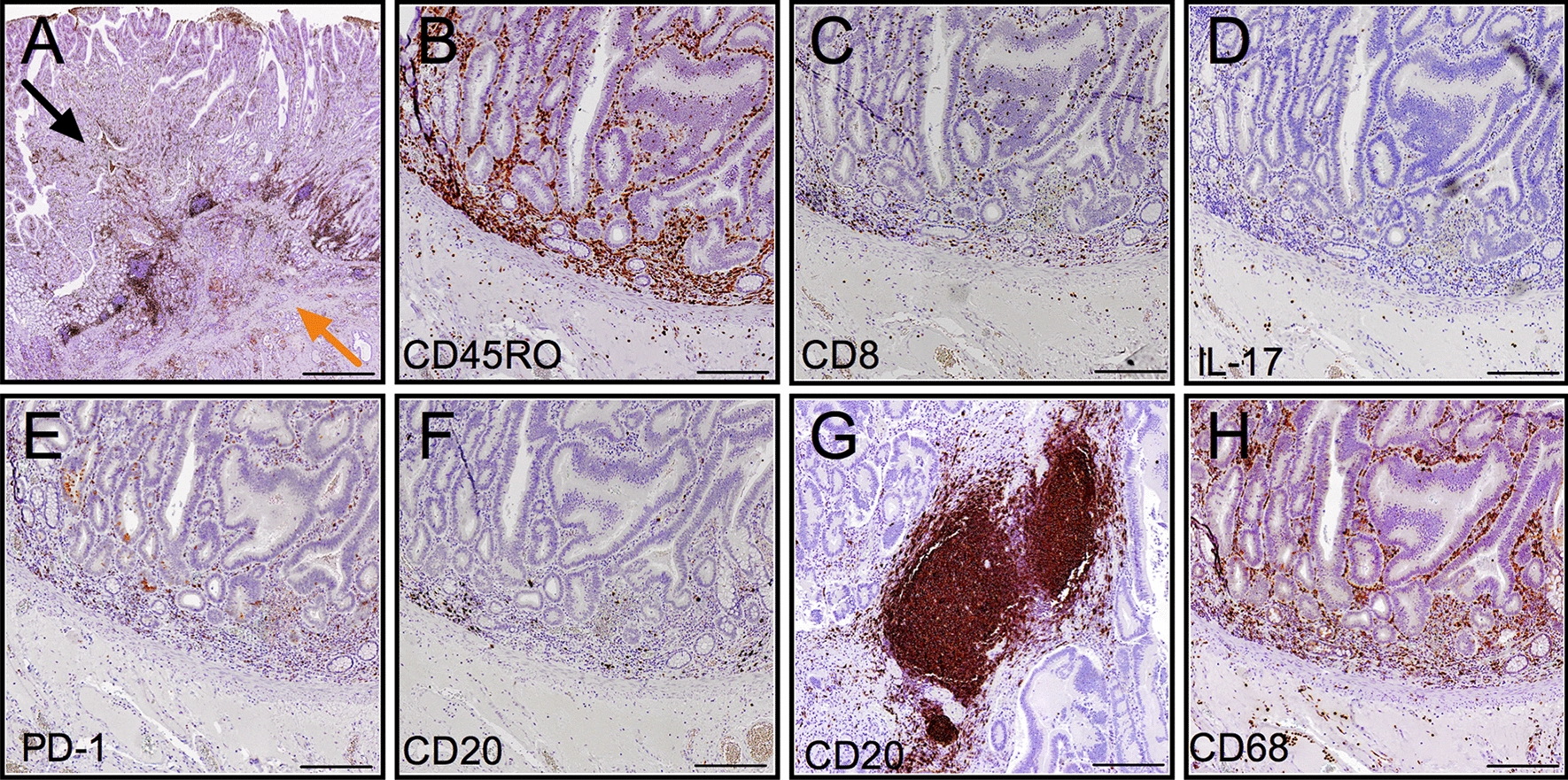
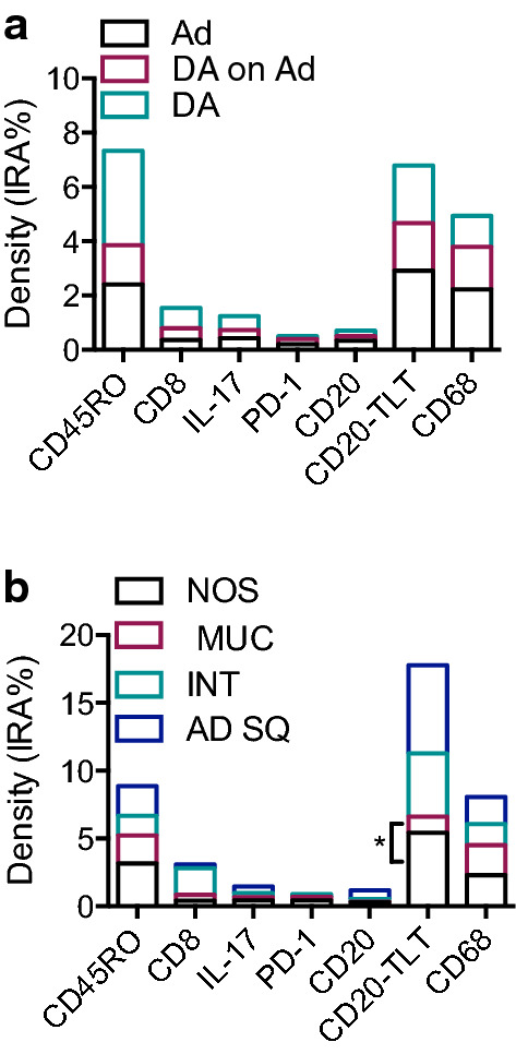
Tissue slides from paraffin-embedded tumor specimens of 15 consecutive DA and 3 adenomas that underwent a pancreaticoduodenectomy in our center between 2010 to 2018 were immunohistochemically stained. The density (percentage of immune reactive area, IRA%) of immune markers CD45RO, CD8, CD20, IL-17, PD-1, CD68 was quantified by computer-assisted image analysis. Demographic, clinical, histopathological data were collected.
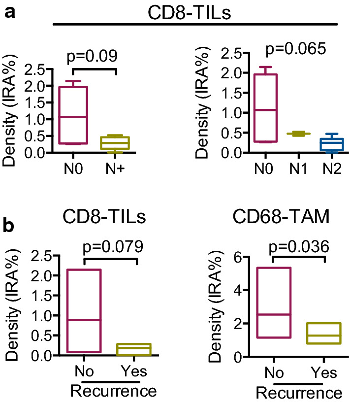
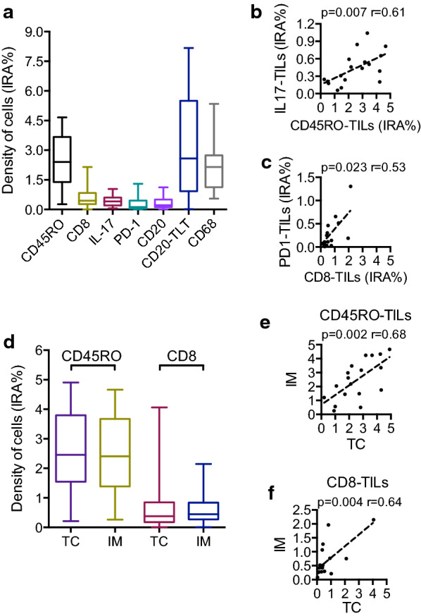
In our population, median IRA % (IQR) of immune subsets was respectively CD45RO-TILs 2.19 (2.14), CD8-TIL 0.42 (0.81), CD20-TILs 0.22 (0.51), CD20-TLT 2.84 (4.64), CD68-TAM 2.19 (1.56), IL17 cells 0.39 (0.39), PD1-TILs 0.19 (0.41). The median follow-up was 47.5 (22.4-63.3) months. At statistical analysis, the density of CD8-TILs inversely correlated with lymph node ratio (p = 0.013), number of metastatic lymph nodes (p = 0.019), and was lower in N+ adenocarcinomas compared to N0 (1.07 vs 0.29; p = 0.093), albeit not significantly. Stratifying patients for the N status, the density of CD8-TILs decreased with the increasing of the N stage (p = 0.065) and was lower in patients who experienced recurrence and died for the disease (0.276 vs 0.641; p = 0.044). Notably, also CD68-TAM distribution was different in patients who had recurrence versus patients who did not (1.028 vs 2.276; p = 0.036).
Immune cells showed variable expression in correlation with common prognostic factors, suggesting T cell infiltration may play a protective role towards lymphatic spread of disease and nodal metastatization. Furthermore, T cell density and macrophage infiltration were associated to a lower risk of recurrence and disease related death. A multicentric approach may be indicated to allow analysis of larger cohorts of patients, potentially increasing the power of our observations.
十二指肠腺癌(DA)是一种罕见但侵袭性强的恶性肿瘤,在过去几十年中发病率不断上升。其低发病率阻碍了对该疾病发病机制及其生物学特性的深入了解,限制了针对性治疗方案的确定。大量证据清楚地表明了免疫细胞在实体瘤中的临床相关性,将免疫特征与手术后的预后相关联。本研究的目的是分析在我们机构接受手术切除的一组十二指肠腺癌中的免疫微环境,并确定其与临床变量的相关性。
对2010年至2018年期间在我们中心接受胰十二指肠切除术的15例连续的DA和3例腺瘤的石蜡包埋肿瘤标本制作的组织切片进行免疫组织化学染色。通过计算机辅助图像分析对免疫标志物CD45RO、CD8、CD20、IL-17、PD-1、CD68的密度(免疫反应面积百分比,IRA%)进行量化。收集人口统计学、临床、组织病理学数据。
在我们的研究人群中,免疫亚群的中位IRA%(四分位间距)分别为:CD45RO-TILs 2.19(2.14)、CD8-TIL 0.42(0.81)、CD20-TILs 0.22(0.51)、CD20-TLT 2.84(4.64)、CD68-TAM 2.19(1.56)、IL17细胞0.39(0.39)、PD1-TILs 0.19(0.41)。中位随访时间为47.5(22.4 - 63.3)个月。经统计学分析,CD8-TILs的密度与淋巴结比率呈负相关(p = 0.013),与转移淋巴结数量呈负相关(p = 0.019),且N +腺癌中的CD8-TILs密度低于N0腺癌(分别为1.07对0.29;p = 0.093),尽管差异不显著。根据N状态对患者进行分层,CD8-TILs的密度随着N分期的增加而降低(p = 0.065),在经历复发并死于该疾病的患者中较低(0.276对0.641;p = 0.044)。值得注意的是,复发患者与未复发患者的CD68-TAM分布也不同(1.028对2.276;p = 0.036)。
免疫细胞显示出与常见预后因素相关的可变表达,提示T细胞浸润可能对疾病的淋巴扩散和淋巴结转移起保护作用。此外,T细胞密度和巨噬细胞浸润与较低的复发风险和疾病相关死亡风险相关。可能需要采用多中心研究方法来分析更大规模的患者队列,从而可能增强我们观察结果的说服力。