Wang Qing Jun, Jung Kyung Sik, Mohan Kabhilan, Kleinman Mark E
Department of Ophthalmology and Visual Sciences, University of Kentucky, Lexington, KY, United States.
Markey Cancer Center, University of Kentucky, Lexington, KY, United States.
Data Brief. 2020 Jul 25;32:106076. doi: 10.1016/j.dib.2020.106076. eCollection 2020 Oct.
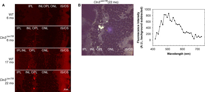
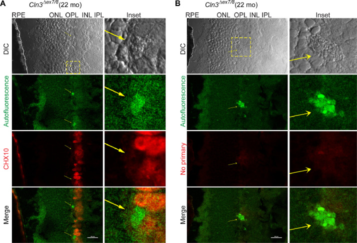
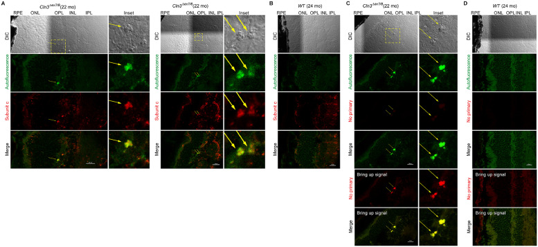
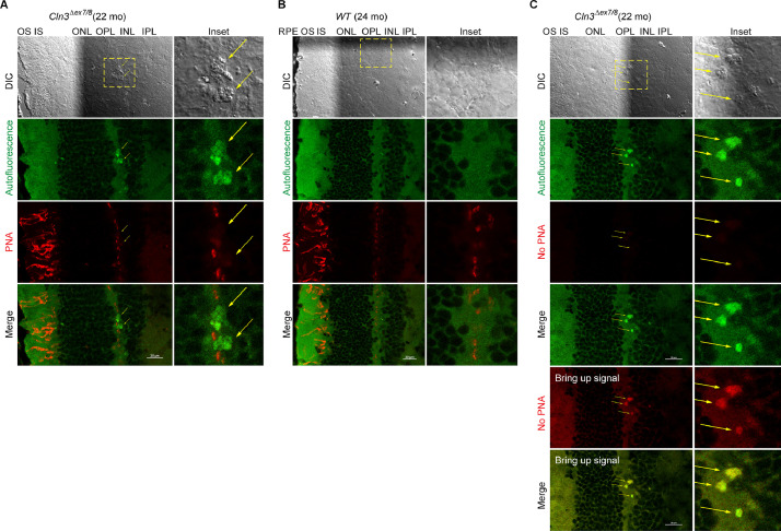
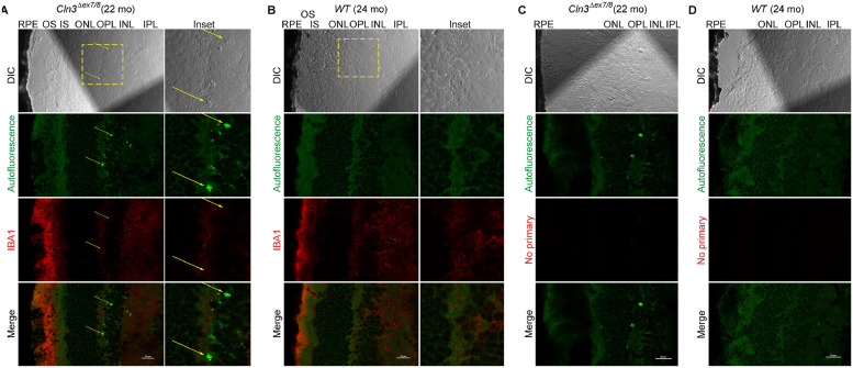
Juvenile neuronal ceroid lipofuscinosis (JNCL, . juvenile Batten disease or CLN3 disease), a lethal pediatric neurodegenerative disease without cure, often presents with vision impairment and characteristic ophthalmoscopic features including focal areas of hyper-autofluorescence. In the associated research article "Loss of , the gene mutated in juvenile neuronal ceroid lipofuscinosis, leads to metabolic impairment and autophagy induction in retinal pigment epithelium" (Zhong et al., 2020) [1], we reported ophthalmoscopic observations of focal autofluorescent lesions or puncta in the mouse retina at as young as 8 month old. In this data article, we performed differential interference contrast and confocal imaging analyses in all retinal layers to localize and characterize these autofluorescent lesions, including their spectral characteristics and morphology. We further studied colocalization of these autofluorescent lesions with the JNCL marker mitochondrial ATP synthase F0 sub-complex subunit C and various established retinal cell type markers.
青少年神经元蜡样脂褐质沉积症(JNCL,即青少年型巴滕病或CLN3病)是一种致命的儿童神经退行性疾病,目前无法治愈,常表现为视力损害以及特征性的眼底镜表现,包括局灶性高自发荧光区域。在相关研究文章《青少年神经元蜡样脂褐质沉积症中发生突变的 基因缺失导致视网膜色素上皮细胞代谢受损和自噬诱导》(钟等人,2020年)[1]中,我们报告了早在8个月大的小鼠视网膜中就观察到局灶性自发荧光病变或斑点的眼底镜检查结果。在这篇数据文章中,我们对所有视网膜层进行了微分干涉对比和共聚焦成像分析,以定位和表征这些自发荧光病变,包括它们的光谱特征和形态。我们进一步研究了这些自发荧光病变与JNCL标志物线粒体ATP合酶F0亚复合体亚基C以及各种已确定的视网膜细胞类型标志物的共定位情况。