Institute of Oral Biology, Faculty of Dentistry, University of Oslo, Oslo, Norway.
Department of Medical Biochemistry, Blood Cell Research Group, Oslo University Hospital, Ullevål, Oslo, Norway.
PLoS One. 2020 Sep 4;15(9):e0238591. doi: 10.1371/journal.pone.0238591. eCollection 2020.
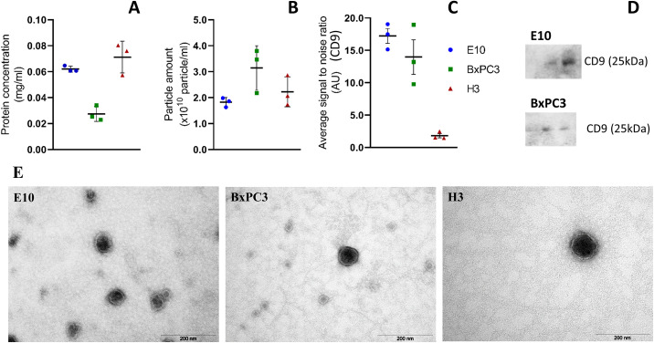
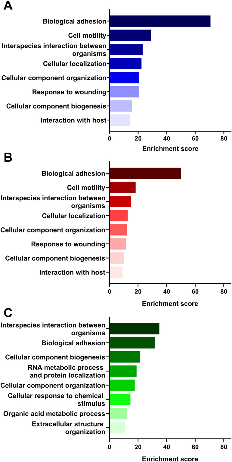
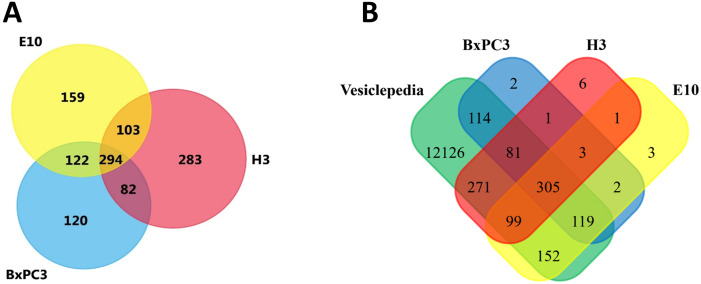
Extracellular vesicles (EVs), are important for intercellular communication in both physiological and pathological processes. To explore the potential of cancer derived EVs as disease biomarkers for diagnosis, monitoring, and treatment decision, it is necessary to thoroughly characterize their biomolecular content. The aim of the study was to characterize and compare the protein content of EVs derived from three different cancer cell lines in search of a specific molecular signature, with emphasis on proteins related to the carcinogenic process. Oral squamous cell carcinoma (OSCC), pancreatic ductal adenocarcinoma (PDAC) and melanoma brain metastasis cell lines were cultured in CELLine AD1000 flasks. EVs were isolated by ultrafiltration and size-exclusion chromatography and characterized. Next, the isolated EVs underwent liquid chromatography-mass spectrometry (LC-MS) analysis for protein identification. Functional enrichment analysis was performed for a more general overview of the biological processes involved. More than 600 different proteins were identified in EVs from each particular cell line. Here, 14%, 10%, and 24% of the identified proteins were unique in OSCC, PDAC, and melanoma vesicles, respectively. A specific protein profile was discovered for each cell line, e.g., EGFR in OSCC, Muc5AC in PDAC, and FN1 in melanoma vesicles. Nevertheless, 25% of all the identified proteins were common to all cell lines. Functional enrichment analysis linked the proteins in each data set to biological processes such as "biological adhesion", "cell motility", and "cellular component biogenesis". EV proteomics discovered cancer-specific protein profiles, with proteins involved in processes promoting tumor progression. In addition, the biological processes associated to the melanoma-derived EVs were distinct from the ones linked to the EVs isolated from OSCC and PDAC. The malignancy specific biomolecular cues in EVs may have potential applications as diagnostic biomarkers and in therapy.
细胞外囊泡 (EVs) 在生理和病理过程中的细胞间通讯中起着重要作用。为了探索源自癌症的 EVs 作为疾病生物标志物用于诊断、监测和治疗决策的潜力,有必要彻底描述其生物分子含量。本研究的目的是表征和比较三种不同癌细胞系来源的 EVs 的蛋白质含量,以寻找特定的分子特征,重点是与致癌过程相关的蛋白质。口腔鳞状细胞癌 (OSCC)、胰腺导管腺癌 (PDAC) 和黑色素瘤脑转移细胞系在 CELLine AD1000 培养瓶中培养。通过超滤和大小排阻色谱法分离 EVs 并进行表征。然后,对分离的 EVs 进行液相色谱-质谱 (LC-MS) 分析以鉴定蛋白质。进行功能富集分析以更全面地了解所涉及的生物学过程。从每个特定细胞系的 EV 中鉴定出超过 600 种不同的蛋白质。在这里,在 OSCC、PDAC 和黑色素瘤囊泡中分别有 14%、10%和 24%的鉴定蛋白是独特的。为每个细胞系发现了特定的蛋白质谱,例如,OSCC 中的 EGFR、PDAC 中的 Muc5AC 和黑色素瘤囊泡中的 FN1。然而,所有鉴定蛋白的 25%在所有细胞系中是共同的。功能富集分析将每个数据集的蛋白质与生物学过程联系起来,例如“生物粘附”、“细胞运动”和“细胞成分生物发生”。EV 蛋白质组学发现了癌症特异性的蛋白质谱,其中涉及促进肿瘤进展的过程的蛋白质。此外,与从 OSCC 和 PDAC 分离的 EV 相关的生物学过程与从黑色素瘤衍生的 EV 相关的生物学过程不同。EV 中恶性肿瘤特异性生物分子线索可能具有作为诊断生物标志物和治疗的潜在应用。