• 文献检索
  • 文档翻译
  • 深度研究
  • 学术资讯
  • Suppr Zotero 插件Zotero 插件
  • 邀请有礼
  • 套餐&价格
  • 历史记录
应用&插件
Suppr Zotero 插件Zotero 插件浏览器插件Mac 客户端Windows 客户端微信小程序
定价
高级版会员购买积分包购买API积分包
服务
文献检索文档翻译深度研究API 文档MCP 服务
关于我们
关于 Suppr公司介绍联系我们用户协议隐私条款
关注我们

Suppr 超能文献

核心技术专利:CN118964589B侵权必究
粤ICP备2023148730 号-1Suppr @ 2026

文献检索

告别复杂PubMed语法,用中文像聊天一样搜索,搜遍4000万医学文献。AI智能推荐,让科研检索更轻松。

立即免费搜索

文件翻译

保留排版,准确专业,支持PDF/Word/PPT等文件格式,支持 12+语言互译。

免费翻译文档

深度研究

AI帮你快速写综述,25分钟生成高质量综述,智能提取关键信息,辅助科研写作。

立即免费体验

基于间充质干细胞的 Smad7 基因治疗实验性肝硬变。

Mesenchymal stem cell-based Smad7 gene therapy for experimental liver cirrhosis.

机构信息

Department of Infectious Diseases, Shenzhen People's Hospital (The Second Clinical Medical College, Jinan University; The First Affiliated Hospital, Southern University of Science and Technology), 1017 Dong Men Bei Road, Luo Hu District, Shenzhen, 518020, Guangdong Province, People's Republic of China.

Centre for Atherothrombosis and Metabolic Disease, Hull York Medical School, University of Hull, Hull, HU6 7RX, UK.

出版信息

Stem Cell Res Ther. 2020 Sep 14;11(1):395. doi: 10.1186/s13287-020-01911-4.

DOI:10.1186/s13287-020-01911-4
PMID:32928296
原文链接:https://pmc.ncbi.nlm.nih.gov/articles/PMC7489041/
Abstract

BACKGROUND

Bone mesenchymal stem cells (MSCs) can promote liver regeneration and inhibit inflammation and hepatic fibrosis. MSCs also can serve as a vehicle for gene therapy. Smad7 is an essential negative regulatory gene in the TGF-β1/Smad signalling pathway. Activation of TGF-β1/Smad signalling accelerates liver inflammation and fibrosis; we therefore hypothesized that MSCs overexpressing the Smad7 gene might be a new cell therapy approach for treating liver fibrosis via the inhibition of TGF-β1/Smad signalling.

METHODS

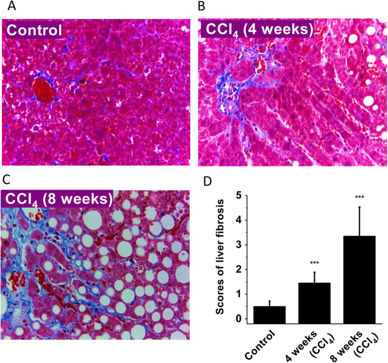
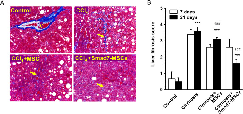
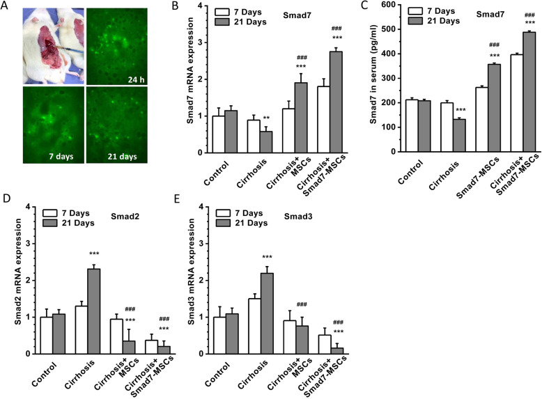
MSCs were isolated from 6-week-old Wistar rats and transduced with the Smad7 gene using a lentivirus vector. Liver cirrhosis was induced by subcutaneous injection of carbon tetrachloride (CCl) for 8 weeks. The rats with established liver cirrhosis were treated with Smad7-MSCs by direct injection of cells into the main lobes of the liver. The expression of Smad7, Smad2/3 and fibrosis biomarkers or extracellular matrix proteins and histopathological change were assessed by quantitative PCR, ELISA and Western blotting and staining.

RESULTS

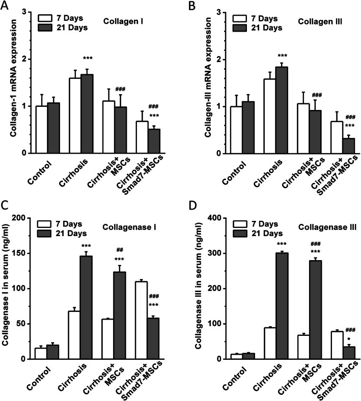
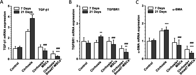
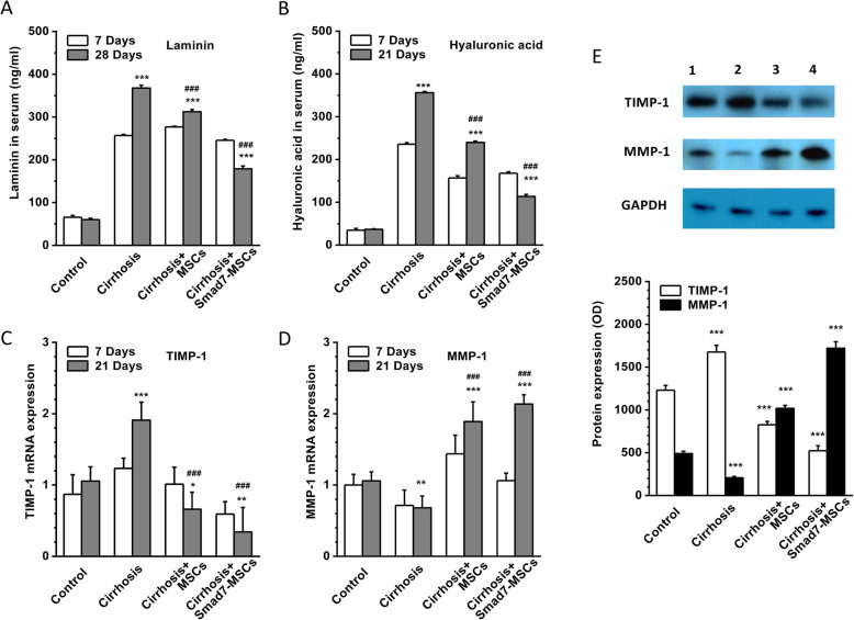
The mRNA and protein level of Smad7 in the recipient liver and serum were increased after treating with Smad-MSCs for 7 and 21 days (P < 0.001). The serum levels of collagen I and III and collagenase I and III were significantly (P < 0.001) reduced after the treatment with Smad7-MSCs. The mRNA levels of TGF-β1, TGFBR1, α-SMA, TIMP-1, laminin and hyaluronic acid were decreased (P < 0.001), while MMP-1 increased (P < 0.001). The liver fibrosis score and liver function were significantly alleviated after the cell therapy.

CONCLUSIONS

The findings suggest that the MSC therapy with Smad7-MSCs is effective in the treatment of liver fibrosis in the CCl-induced liver cirrhosis model. Inhibition of TGF-β1 signalling pathway by enhancement of Smad-7 expression could be a feasible cell therapy approach to mitigate liver cirrhosis.

摘要

背景

骨髓间充质干细胞(MSCs)可促进肝脏再生,抑制炎症和肝纤维化。MSCs 还可以作为基因治疗的载体。Smad7 是 TGF-β1/Smad 信号通路中的一个重要负调控基因。TGF-β1/Smad 信号的激活会加速肝脏炎症和纤维化;因此,我们假设过表达 Smad7 基因的 MSCs 可能通过抑制 TGF-β1/Smad 信号通路成为治疗肝纤维化的一种新的细胞治疗方法。

方法

从 6 周龄 Wistar 大鼠中分离 MSCs,并使用慢病毒载体转染 Smad7 基因。通过皮下注射四氯化碳(CCl)8 周诱导肝硬化。用细胞直接注射到肝脏主要叶来治疗 Smad7-MSCs 诱导的肝硬化大鼠。通过定量 PCR、ELISA 和 Western 印迹及染色评估 Smad7、Smad2/3 和纤维化生物标志物或细胞外基质蛋白的表达和组织病理学变化。

结果

Smad7-MSCs 治疗 7 和 21 天后,受体肝和血清中 Smad7 的 mRNA 和蛋白水平均升高(P<0.001)。Smad7-MSCs 治疗后,血清中 I 型和 III 型胶原及胶原酶 I 和 III 的水平显著降低(P<0.001)。TGF-β1、TGFBR1、α-SMA、TIMP-1、层粘连蛋白和透明质酸的 mRNA 水平降低(P<0.001),而 MMP-1 增加(P<0.001)。细胞治疗后,肝纤维化评分和肝功能明显改善。

结论

研究结果表明,MSC 疗法联合 Smad7-MSCs 对 CCl 诱导的肝硬化模型中的肝纤维化治疗有效。通过增强 Smad-7 表达抑制 TGF-β1 信号通路可能是减轻肝硬化的可行细胞治疗方法。

https://cdn.ncbi.nlm.nih.gov/pmc/blobs/575c/7489041/bbadae712d39/13287_2020_1911_Fig6_HTML.jpg
https://cdn.ncbi.nlm.nih.gov/pmc/blobs/575c/7489041/9150766041b9/13287_2020_1911_Fig1_HTML.jpg
https://cdn.ncbi.nlm.nih.gov/pmc/blobs/575c/7489041/018d049f8b22/13287_2020_1911_Fig2_HTML.jpg
https://cdn.ncbi.nlm.nih.gov/pmc/blobs/575c/7489041/f99f06ba5c49/13287_2020_1911_Fig3_HTML.jpg
https://cdn.ncbi.nlm.nih.gov/pmc/blobs/575c/7489041/c0acbe431fb1/13287_2020_1911_Fig4_HTML.jpg
https://cdn.ncbi.nlm.nih.gov/pmc/blobs/575c/7489041/72b9a77fa21e/13287_2020_1911_Fig5_HTML.jpg
https://cdn.ncbi.nlm.nih.gov/pmc/blobs/575c/7489041/bbadae712d39/13287_2020_1911_Fig6_HTML.jpg

相似文献

1
Mesenchymal stem cell-based Smad7 gene therapy for experimental liver cirrhosis.基于间充质干细胞的 Smad7 基因治疗实验性肝硬变。
Stem Cell Res Ther. 2020 Sep 14;11(1):395. doi: 10.1186/s13287-020-01911-4.
2
[Dynamic expression of TGF-beta1/Smad protein in CCl4-induced liver fibrosis and its significance in rats].[转化生长因子β1/Smad蛋白在四氯化碳诱导的大鼠肝纤维化中的动态表达及其意义]
Zhonghua Shi Yan He Lin Chuang Bing Du Xue Za Zhi. 2011 Oct;25(5):334-7.
3
Shugan Huoxue Huayu Fang attenuates carbon tetrachloride-induced hepatic fibrosis in rats by inhibiting transforming growth factor-β1/Smad signaling.疏肝活血化瘀方通过抑制转化生长因子-β1/Smad信号通路减轻四氯化碳诱导的大鼠肝纤维化。
J Tradit Chin Med. 2022 Feb;42(1):65-72. doi: 10.19852/j.cnki.jtcm.20210624.001.
4
Effect of Human Umbilical Cord Mesenchymal Stem Cells Transfected with HGF on TGF-β1/Smad Signaling Pathway in Carbon Tetrachloride-Induced Liver Fibrosis Rats.人脐带间充质干细胞转染 HGF 对四氯化碳致肝纤维化大鼠 TGF-β1/Smad 信号通路的影响。
Stem Cells Dev. 2020 Nov 1;29(21):1395-1406. doi: 10.1089/scd.2020.0060. Epub 2020 Sep 24.
5
Inhibitory Effect of Corilagin on miR-21-Regulated Hepatic Fibrosis Signaling Pathway.鞣花酸对 miR-21 调控的肝纤维化信号通路的抑制作用。
Am J Chin Med. 2019;47(7):1541-1569. doi: 10.1142/S0192415X19500794.
6
Coexpression of Smad7 and UPA attenuates carbon tetrachloride-induced rat liver fibrosis.Smad7 和 UPA 的共表达可减轻四氯化碳诱导的大鼠肝纤维化。
Med Sci Monit. 2012 Oct;18(10):BR394-401. doi: 10.12659/msm.883479.
7
Effect of Function-Enhanced Mesenchymal Stem Cells Infected With Decorin-Expressing Adenovirus on Hepatic Fibrosis.感染表达核心蛋白聚糖腺病毒的功能增强间充质干细胞对肝纤维化的影响
Stem Cells Transl Med. 2016 Sep;5(9):1247-56. doi: 10.5966/sctm.2015-0323. Epub 2016 Jun 30.
8
Smad7-overexpressing rat BMSCs inhibit the fibrosis of hepatic stellate cells by regulating the TGF-β1/Smad signaling pathway.过表达Smad7的大鼠骨髓间充质干细胞通过调节TGF-β1/Smad信号通路抑制肝星状细胞纤维化。
Exp Ther Med. 2017 Sep;14(3):2568-2576. doi: 10.3892/etm.2017.4836. Epub 2017 Jul 25.
9
Histone deacetylase inhibitor suberoylanilide hydroxamic acid alleviates liver fibrosis by suppressing the transforming growth factor-β1 signal pathway.组蛋白去乙酰化酶抑制剂丁酸钠通过抑制转化生长因子-β1 信号通路缓解肝纤维化。
Hepatobiliary Pancreat Dis Int. 2018 Oct;17(5):423-429. doi: 10.1016/j.hbpd.2018.09.013. Epub 2018 Sep 15.
10
Puerarin, isolated from Pueraria lobata (Willd.), protects against hepatotoxicity via specific inhibition of the TGF-β1/Smad signaling pathway, thereby leading to anti-fibrotic effect.葛根素从野葛(Willd.)中分离得到,通过特异性抑制 TGF-β1/Smad 信号通路来保护肝脏免受毒性损伤,从而发挥抗纤维化作用。
Phytomedicine. 2013 Oct 15;20(13):1172-9. doi: 10.1016/j.phymed.2013.06.008. Epub 2013 Jul 13.

引用本文的文献

1
Overexpression of Decorin Optimizes the Treatment Efficacy of Umbilical Cord Mesenchymal Stem Cells in Bleomycin-Induced Pulmonary Fibrosis in Rats.核心蛋白聚糖的过表达优化了脐带间充质干细胞对博莱霉素诱导的大鼠肺纤维化的治疗效果。
Stem Cells Int. 2025 May 21;2025:6324980. doi: 10.1155/sci/6324980. eCollection 2025.
2
Application of mesenchymal stem cells in liver fibrosis and regeneration.间充质干细胞在肝纤维化和肝再生中的应用。
Liver Res. 2024 Nov 26;8(4):246-258. doi: 10.1016/j.livres.2024.11.004. eCollection 2024 Dec.
3
Airway stenosis: classification, pathogenesis, and clinical management.

本文引用的文献

1
The global, regional, and national burden of cirrhosis by cause in 195 countries and territories, 1990-2017: a systematic analysis for the Global Burden of Disease Study 2017.2017 年全球疾病负担研究:1990-2017 年 195 个国家和地区按病因划分的肝硬化全球、区域和国家负担:系统分析。
Lancet Gastroenterol Hepatol. 2020 Mar;5(3):245-266. doi: 10.1016/S2468-1253(19)30349-8. Epub 2020 Jan 22.
2
TGF-β orchestrates fibrogenic and developmental EMTs via the RAS effector RREB1.TGF-β 通过 RAS 效应因子 RREB1 来协调纤维化和发育性 EMT。
Nature. 2020 Jan;577(7791):566-571. doi: 10.1038/s41586-019-1897-5. Epub 2020 Jan 8.
3
气道狭窄:分类、发病机制及临床管理
MedComm (2020). 2025 Jan 26;6(2):e70076. doi: 10.1002/mco2.70076. eCollection 2025 Feb.
4
Cell therapy for end-stage liver disease: Current state and clinical challenge.终末期肝病的细胞治疗:现状与临床挑战
Chin Med J (Engl). 2024 Dec 5;137(23):2808-2820. doi: 10.1097/CM9.0000000000003332. Epub 2024 Nov 18.
5
Overexpressing Adipose-Derived Mesenchymal Stem Cells Mitigate Alcohol-Induced Liver Damage and Gut Permeability.过表达脂肪间充质干细胞减轻酒精性肝损伤和肠道通透性。
Int J Mol Sci. 2024 Aug 4;25(15):8509. doi: 10.3390/ijms25158509.
6
Effectiveness and mechanisms of mesenchymal stem cell therapy in preclinical animal models of hepatic fibrosis: a systematic review and meta-analysis.间充质干细胞疗法在肝纤维化临床前动物模型中的有效性及机制:一项系统评价和荟萃分析
Front Bioeng Biotechnol. 2024 Jul 22;12:1424253. doi: 10.3389/fbioe.2024.1424253. eCollection 2024.
7
Mesenchymal stem/stromal cells armored by FGF21 ameliorate alcohol-induced liver injury through modulating polarization of macrophages.成纤维细胞生长因子 21 包裹的间充质干细胞/基质细胞通过调节巨噬细胞极化改善酒精性肝损伤。
Hepatol Commun. 2024 Mar 29;8(4). doi: 10.1097/HC9.0000000000000410. eCollection 2024 Apr 1.
8
Cell mediated ECM-degradation as an emerging tool for anti-fibrotic strategy.细胞介导的细胞外基质降解作为一种新兴的抗纤维化策略工具。
Cell Regen. 2023 Sep 1;12(1):29. doi: 10.1186/s13619-023-00172-9.
9
Potential advantages of genetically modified mesenchymal stem cells in the treatment of acute and chronic liver diseases.基因修饰间充质干细胞在治疗急性和慢性肝病中的潜在优势。
Stem Cell Res Ther. 2023 May 24;14(1):138. doi: 10.1186/s13287-023-03364-x.
10
Mesenchymal stromal cells in hepatic fibrosis/cirrhosis: from pathogenesis to treatment.肝纤维化/肝硬化中的间充质基质细胞:从发病机制到治疗。
Cell Mol Immunol. 2023 Jun;20(6):583-599. doi: 10.1038/s41423-023-00983-5. Epub 2023 Feb 24.
Effects of Bone Marrow-Derived Mesenchymal Stem Cells on Hypoxia and the Transforming Growth Factor beta 1 (TGFβ-1) and SMADs Pathway in a Mouse Model of Cirrhosis.
骨髓间充质干细胞对肝硬化小鼠模型缺氧及转化生长因子β1(TGFβ-1)和 SMADs 通路的影响。
Med Sci Monit. 2019 Sep 24;25:7182-7190. doi: 10.12659/MSM.916428.
4
Mesenchymal stem cell therapies for liver cirrhosis: MSCs as "conducting cells" for improvement of liver fibrosis and regeneration.用于肝硬化的间充质干细胞疗法:间充质干细胞作为改善肝纤维化和再生的“传导细胞”
Inflamm Regen. 2019 Sep 9;39:18. doi: 10.1186/s41232-019-0107-z. eCollection 2019.
5
Chronic hepatitis B virus infection.慢性乙型肝炎病毒感染。
Lancet. 2018 Nov 24;392(10161):2313-2324. doi: 10.1016/S0140-6736(18)31865-8.
6
Recognizing the Molecular Multifunctionality and Interactome of TIMP-1.认识 TIMP-1 的分子多功能性和相互作用组。
Trends Cell Biol. 2019 Jan;29(1):6-19. doi: 10.1016/j.tcb.2018.08.006. Epub 2018 Sep 19.
7
Comparison of Two Protocols of Carbon Tetrachloride-Induced Cirrhosis in Rats - Improving Yield and Reproducibility.两种四氯化碳诱导大鼠肝硬化模型方案的比较——提高产量和重现性。
Sci Rep. 2018 Jun 15;8(1):9163. doi: 10.1038/s41598-018-27427-9.
8
Chronic Hepatitis B Infection: A Review.慢性乙型肝炎感染:综述。
JAMA. 2018 May 1;319(17):1802-1813. doi: 10.1001/jama.2018.3795.
9
Contextual determinants of TGFβ action in development, immunity and cancer.转化生长因子β(TGFβ)在发育、免疫和癌症中作用的情境决定因素。
Nat Rev Mol Cell Biol. 2018 Jul;19(7):419-435. doi: 10.1038/s41580-018-0007-0.
10
A pooled analysis of mesenchymal stem cell-based therapy for liver disease.基于间充质干细胞的肝病治疗的荟萃分析。
Stem Cell Res Ther. 2018 Mar 21;9(1):72. doi: 10.1186/s13287-018-0816-2.