Institute of Muscle Biology and Growth, Growth and Development Unit, Leibniz Institute for Farm Animal Biology (FBN), Wilhelm-Stahl-Allee 2, 18196, Dummerstorf, Germany.
Research Group Stem Cells in Regeneration of Skeletal Muscle, Leibniz Institute on Aging, 07745, Jena, Germany.
In Vitro Cell Dev Biol Anim. 2020 Sep;56(8):585-592. doi: 10.1007/s11626-020-00492-z. Epub 2020 Sep 22.
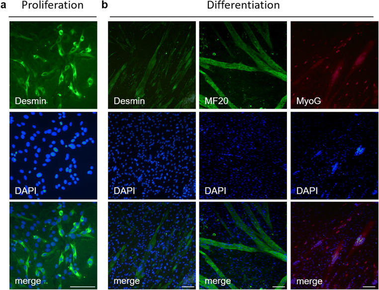
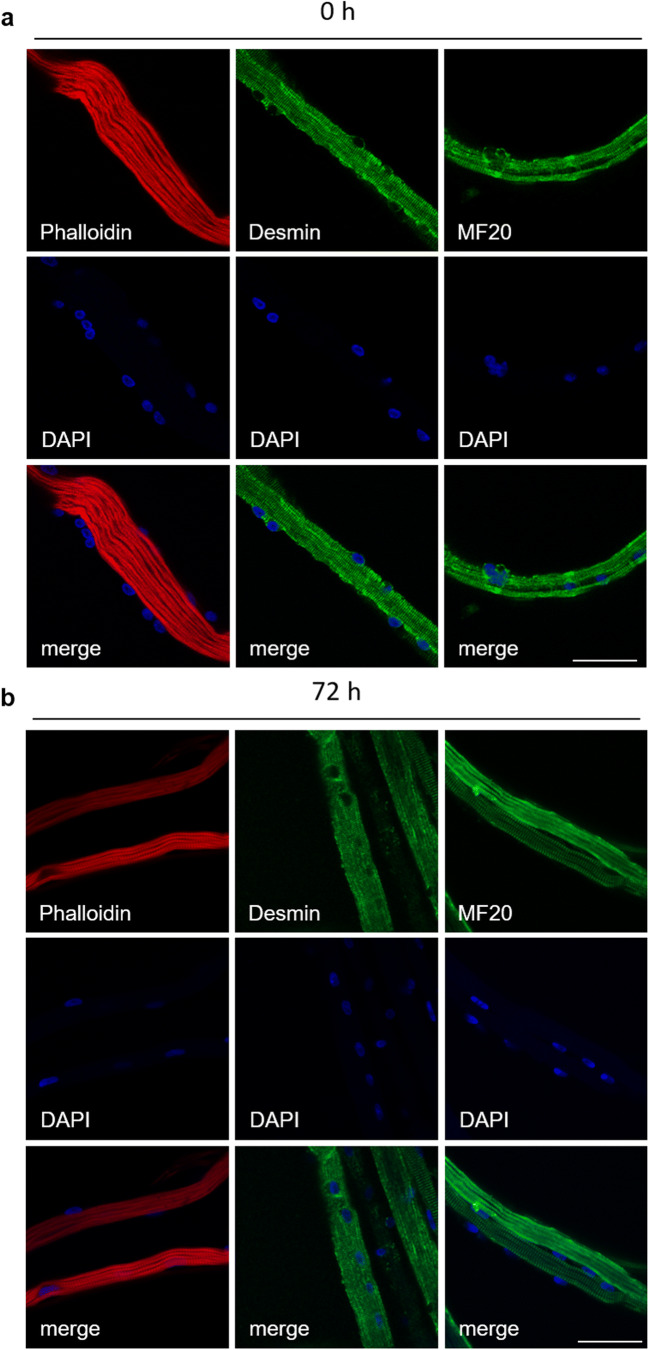
The isolation and cultivation of intact, single myofibers presents a superior approach for studying myogenic cells in their native position. The cells' characteristics remain more similar to muscle tissue than in cell culture. Nevertheless, no routinely used method in higher vertebrates exists. Therefore, we aimed at establishing the isolation and cultivation of single myofibers from porcine muscle. For the first time, we implemented the isolation of intact myofibers from porcine fibularis tertius muscle by enzymatic digestion and their subsequent cultivation under floating conditions. Confocal microscopy showed intact myofibrill structures in isolated myofibers. Myogenic cells were able to proliferate at their parent myofiber as shown by the increase of myonuclear number during culture. Additionally, the described method can be used to investigate myogenic cells migrated from isolated myofibers. These cells expressed myogenic markers and were able to differentiate. In the future, our method can be used for genetic manipulation of cells at myofibers, investigation of growth factors or pharmacological substances, and determination of interactions between myofibers and associated cells. Working with isolated myofibers has the potential to bridge conventional cell culture and animal experiments. Adapting the method to porcine muscle allows for application possibilities in veterinary medicine as well as in biomedical research, which cannot be addressed in rodent model systems.
从完整的、单一的肌纤维中分离和培养可以为研究天然位置的成肌细胞提供一种优越的方法。与细胞培养相比,这些细胞的特征更接近肌肉组织。然而,高等脊椎动物中还没有常规使用的方法。因此,我们的目的是建立从猪肌肉中分离和培养单一肌纤维的方法。我们首次通过酶消化从猪腓肠肌中分离出完整的肌纤维,并在漂浮条件下对其进行后续培养。共聚焦显微镜显示分离的肌纤维中存在完整的肌原纤维结构。肌源性细胞能够在其母肌纤维上增殖,这可以通过培养过程中核数的增加来证明。此外,所描述的方法可用于研究从分离的肌纤维中迁移出来的成肌细胞。这些细胞表达成肌细胞标志物,并能够分化。将来,我们的方法可以用于肌纤维上的细胞遗传操作、生长因子或药物物质的研究,以及肌纤维和相关细胞之间相互作用的确定。使用分离的肌纤维具有将传统细胞培养和动物实验联系起来的潜力。将该方法应用于猪肌肉,不仅可以在兽医医学中,也可以在生物医学研究中得到应用,而这些在啮齿动物模型系统中是无法实现的。