Kaneko Fumiya, Edama Mutsuaki, Ikezu Masahiro, Matsuzawa Kanta, Hirabayashi Ryo, Kageyama Ikuo
Institute for Human Movement and Medical Sciences, Niigata University of Health and Welfare, Niigata, Japan.
Department of Anatomy, School of Life Dentistry at Niigata, Nippon Dental University, Niigata, Japan.
Orthop J Sports Med. 2020 Sep 18;8(9):2325967120947725. doi: 10.1177/2325967120947725. eCollection 2020 Sep.
Two types of stress, bending stress and traction stress, have been reported to be involved in the mechanism of Jones fracture. However, little is known about the risk factors for traction stress.
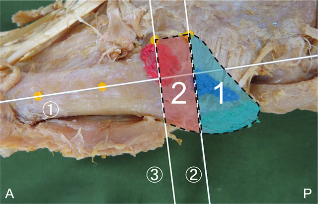
To classify the attachment position of the peroneus brevis muscle (PB), peroneus tertius (PT), lateral band of the plantar aponeurosis (LB), and the long plantar ligament (LPL), focusing on the zone where a Jones fracture occurs (zone 2), and to compare the footprint area of each tissue type.
Descriptive laboratory study.
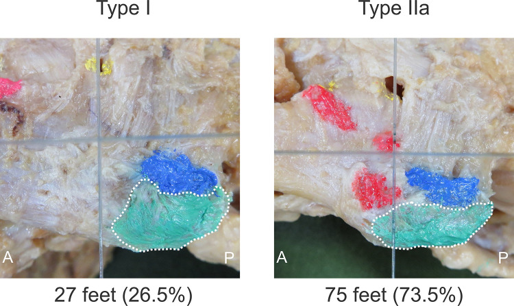
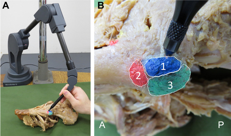
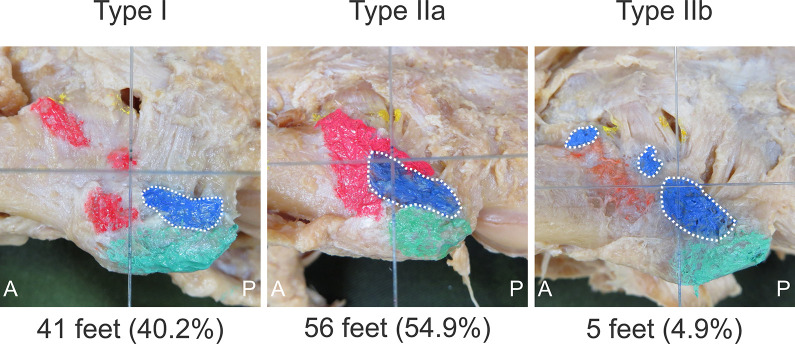
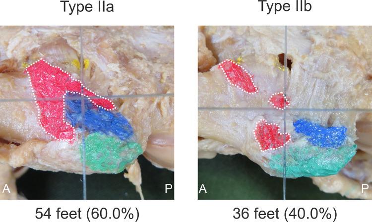
This study examined 102 legs from 55 Japanese cadavers. Type classification was performed by focusing on the positional relationship between each tissue attachment and the zone where Jones fracture occurs (zone 2). The classifications were as follows: type I, attached proximal to the border between zones 1 and 2; type IIa, attached to the border between zones 1 and 2 with one attached part; and type IIb, attached across the border between zones 1 and 2 with two or more attached parts. The footprint areas of the PB, PT, LB, and LPL were compared between tissue types and within each attachment classification.
The PB was recorded as type I in 41 feet (40.2%), type IIa in 56 feet (54.9%), and type IIb in 5 feet (4.9%); the PT was recorded as type IIa in 54 feet (60.0%) and type IIb in 36 feet (40.0%); and the LB was recorded as type I in 27 feet (26.5%) and type IIa in 75 feet (73.5%). The LPL did not attach to the fifth metatarsal bone. No significant difference was found in the footprint area between type I PB and type I LB.
The results indicate that type I, which attaches proximal to zone 2, occurs with PB and LB, and there was no significant difference in the footprint area between them. These findings suggest that type I is involved in traction stress. In the future, biomechanical research based on the results of this study will be necessary.
The results of this study provide basic research for investigating the mechanism of Jones fracture and the cause of delayed healing.
据报道,两种应力,即弯曲应力和牵引应力,参与了琼斯骨折的机制。然而,关于牵引应力的危险因素知之甚少。
对腓骨短肌(PB)、第三腓骨肌(PT)、足底腱膜外侧束(LB)和足底长韧带(LPL)的附着位置进行分类,重点关注琼斯骨折发生的区域(2区),并比较每种组织类型的足迹面积。
描述性实验室研究。
本研究检查了55具日本尸体的102条腿。通过关注每种组织附着与琼斯骨折发生区域(2区)之间的位置关系进行类型分类。分类如下:I型,附着于1区和2区边界近端;IIa型,通过一个附着部分附着于1区和2区边界;IIb型,通过两个或更多附着部分跨越1区和2区边界附着。比较了不同组织类型之间以及每种附着分类内PB、PT、LB和LPL的足迹面积。
PB记录为I型的有41只足(40.2%),IIa型的有56只足(54.9%),IIb型的有5只足(4.9%);PT记录为IIa型的有54只足(60.0%),IIb型的有36只足(40.0%);LB记录为I型的有27只足(26.5%),IIa型的有75只足(73.5%)。LPL未附着于第五跖骨。I型PB和I型LB的足迹面积之间未发现显著差异。
结果表明,附着于2区近端的I型在PB和LB中出现,且它们之间的足迹面积无显著差异。这些发现表明I型与牵引应力有关。未来,有必要基于本研究结果进行生物力学研究。
本研究结果为研究琼斯骨折的机制和延迟愈合的原因提供了基础研究。