• 文献检索
  • 文档翻译
  • 深度研究
  • 学术资讯
  • Suppr Zotero 插件Zotero 插件
  • 邀请有礼
  • 套餐&价格
  • 历史记录
应用&插件
Suppr Zotero 插件Zotero 插件浏览器插件Mac 客户端Windows 客户端微信小程序
定价
高级版会员购买积分包购买API积分包
服务
文献检索文档翻译深度研究API 文档MCP 服务
关于我们
关于 Suppr公司介绍联系我们用户协议隐私条款
关注我们

Suppr 超能文献

核心技术专利:CN118964589B侵权必究
粤ICP备2023148730 号-1Suppr @ 2026

文献检索

告别复杂PubMed语法,用中文像聊天一样搜索,搜遍4000万医学文献。AI智能推荐,让科研检索更轻松。

立即免费搜索

文件翻译

保留排版,准确专业,支持PDF/Word/PPT等文件格式,支持 12+语言互译。

免费翻译文档

深度研究

AI帮你快速写综述,25分钟生成高质量综述,智能提取关键信息,辅助科研写作。

立即免费体验

基于深度学习的增强视盘照相术。

Deep-learning-based enhanced optic-disc photography.

机构信息

Department of Ophthalmology, Seoul National University College of Medicine, Seoul, Korea.

Department of Ophthalmology, Jeju National University Hospital, Jeju-si, Korea.

出版信息

PLoS One. 2020 Oct 1;15(10):e0239913. doi: 10.1371/journal.pone.0239913. eCollection 2020.

DOI:10.1371/journal.pone.0239913
PMID:33002080
原文链接:https://pmc.ncbi.nlm.nih.gov/articles/PMC7529226/
Abstract

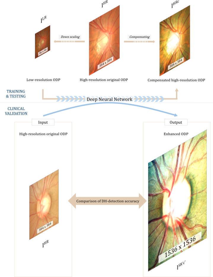
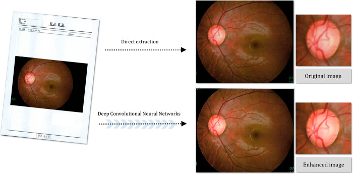
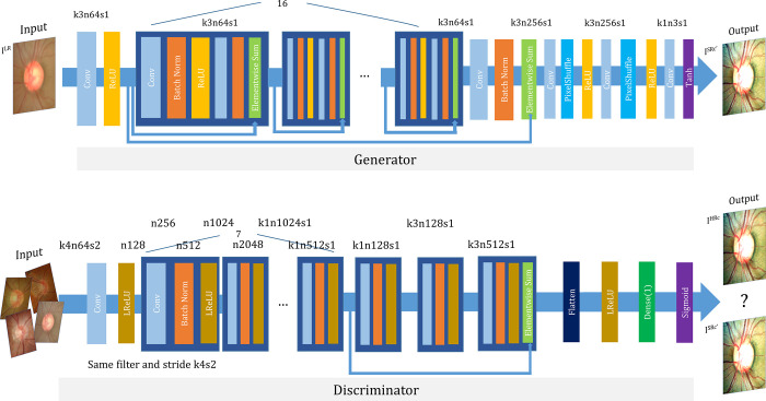
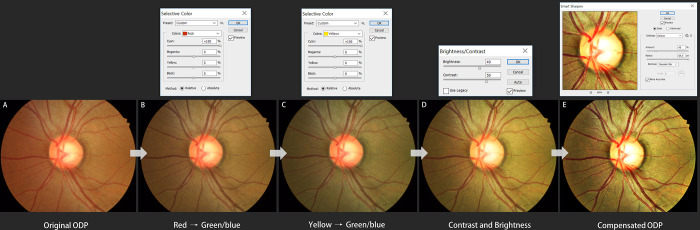
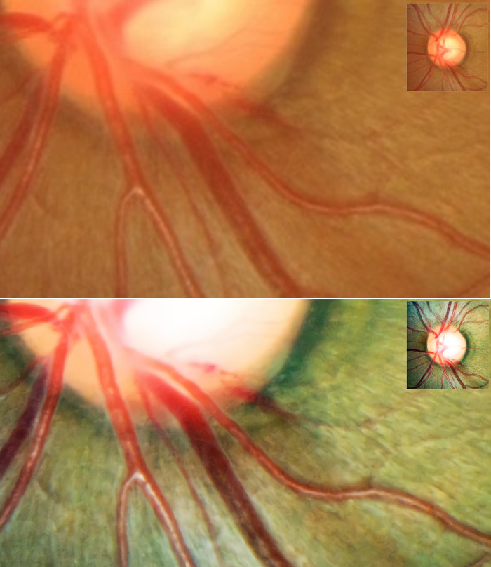
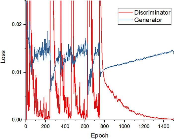
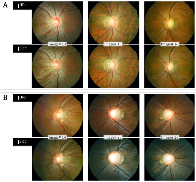
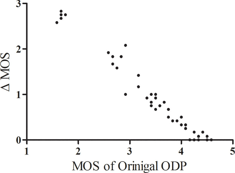
Optic-disc photography (ODP) has proven to be very useful for optic nerve evaluation in glaucoma. In real clinical practice, however, limited patient cooperation, small pupils, or media opacities can limit the performance of ODP. The purpose of this study was to propose a deep-learning approach for increased resolution and improved legibility of ODP by contrast, color, and brightness compensation. Each high-resolution original ODP was transformed into two counterparts: (1) down-scaled 'low-resolution ODPs', and (2) 'compensated high-resolution ODPs' produced via enhancement of the visibility of the optic disc margin and surrounding retinal vessels using a customized image post-processing algorithm. Then, the differences between these two counterparts were directly learned through a super-resolution generative adversarial network (SR-GAN). Finally, by inputting the high-resolution ODPs into SR-GAN, 4-times-up-scaled and overall-color-and-brightness-transformed 'enhanced ODPs' could be obtained. General ophthalmologists were instructed (1) to assess each ODP's image quality, and (2) to note any abnormal findings, at 1-month intervals. The image quality score for the enhanced ODPs was significantly higher than that for the original ODP, and the overall optic disc hemorrhage (DH)-detection accuracy was significantly higher with the enhanced ODPs. We expect that this novel deep-learning approach will be applied to various types of ophthalmic images.

摘要

眼底照相(ODP)已被证明在青光眼视神经评估中非常有用。然而,在实际临床实践中,由于患者配合度有限、瞳孔较小或介质混浊,ODP 的性能可能会受限。本研究旨在提出一种深度学习方法,通过对比度、颜色和亮度补偿来提高 ODP 的分辨率和清晰度。每个高分辨率原始 ODP 都转换为两个副本:(1)降采样的“低分辨率 ODP”,以及(2)通过使用定制的图像后处理算法增强视盘边缘和周围视网膜血管的可视性而生成的“补偿高分辨率 ODP”。然后,通过超分辨率生成对抗网络(SR-GAN)直接学习这两个副本之间的差异。最后,通过将高分辨率 ODP 输入到 SR-GAN 中,可以获得 4 倍放大和整体颜色和亮度变换的“增强 ODP”。普通眼科医生被指示(1)评估每个 ODP 的图像质量,(2)在 1 个月的间隔内注意任何异常发现。增强 ODP 的图像质量评分明显高于原始 ODP,增强 ODP 的总体视盘出血(DH)检测准确率也明显更高。我们期望这种新的深度学习方法将应用于各种类型的眼科图像。

https://cdn.ncbi.nlm.nih.gov/pmc/blobs/1b54/7529226/79df554ad3da/pone.0239913.g008.jpg
https://cdn.ncbi.nlm.nih.gov/pmc/blobs/1b54/7529226/20074b022c4a/pone.0239913.g001.jpg
https://cdn.ncbi.nlm.nih.gov/pmc/blobs/1b54/7529226/5ef289a75b55/pone.0239913.g002.jpg
https://cdn.ncbi.nlm.nih.gov/pmc/blobs/1b54/7529226/b6c23faf112a/pone.0239913.g003.jpg
https://cdn.ncbi.nlm.nih.gov/pmc/blobs/1b54/7529226/37040fc3991f/pone.0239913.g004.jpg
https://cdn.ncbi.nlm.nih.gov/pmc/blobs/1b54/7529226/85b2383b6f67/pone.0239913.g005.jpg
https://cdn.ncbi.nlm.nih.gov/pmc/blobs/1b54/7529226/6d404bf33a3f/pone.0239913.g006.jpg
https://cdn.ncbi.nlm.nih.gov/pmc/blobs/1b54/7529226/5ca3c41ade05/pone.0239913.g007.jpg
https://cdn.ncbi.nlm.nih.gov/pmc/blobs/1b54/7529226/79df554ad3da/pone.0239913.g008.jpg

相似文献

1
Deep-learning-based enhanced optic-disc photography.基于深度学习的增强视盘照相术。
PLoS One. 2020 Oct 1;15(10):e0239913. doi: 10.1371/journal.pone.0239913. eCollection 2020.
2
Combined in-depth, 3D, en face imaging of the optic disc, optic disc pits and optic disc pit maculopathy using swept-source megahertz OCT at 1050 nm.使用1050纳米扫频源兆赫兹光学相干断层扫描技术对视盘、视盘小凹和视盘小凹黄斑病变进行联合深度、三维、正面成像。
Graefes Arch Clin Exp Ophthalmol. 2018 Feb;256(2):289-298. doi: 10.1007/s00417-017-3857-9. Epub 2017 Dec 14.
3
Structural characteristics of the acquired optic disc pit and the rate of progressive retinal nerve fiber layer thinning in primary open-angle glaucoma.原发性开角型青光眼获得性视盘凹陷的结构特征及视网膜神经纤维层渐进性变薄的速率
JAMA Ophthalmol. 2015 Oct;133(10):1151-8. doi: 10.1001/jamaophthalmol.2015.2453.
4
Diagnosis of Glaucoma on Retinal Fundus Images Using Deep Learning: Detection of Nerve Fiber Layer Defect and Optic Disc Analysis.基于深度学习的眼底图像青光眼诊断:神经纤维层缺损检测和视盘分析。
Adv Exp Med Biol. 2020;1213:121-132. doi: 10.1007/978-3-030-33128-3_8.
5
WGAN domain adaptation for the joint optic disc-and-cup segmentation in fundus images.基于 WGAN 的眼底图像视盘-杯联合分割领域自适应方法。
Int J Comput Assist Radiol Surg. 2020 Jul;15(7):1205-1213. doi: 10.1007/s11548-020-02144-9. Epub 2020 May 22.
6
Conversion of Single Optic Disc Photography into 3-Dimensional Image.将单张视盘摄影转换为三维图像。
Ophthalmology. 2018 Dec;125(12):1873. doi: 10.1016/j.ophtha.2018.09.010.
7
Estimating visual field loss from monoscopic optic disc photography using deep learning model.使用深度学习模型从单目视神经盘照相估计视野损失。
Sci Rep. 2020 Dec 3;10(1):21052. doi: 10.1038/s41598-020-78144-1.
8
Computer-assisted planimetry associated with Sturge-Weber syndrome.与斯特奇-韦伯综合征相关的计算机辅助平面测量法。
J Audiov Media Med. 2000 Dec;23(4):149-52. doi: 10.1080/01405110050198591.
9
Computer-aided recognition of myopic tilted optic disc using deep learning algorithms in fundus photography.利用深度学习算法在眼底摄影中对近视性倾斜视盘进行计算机辅助识别。
BMC Ophthalmol. 2020 Oct 9;20(1):407. doi: 10.1186/s12886-020-01657-w.
10
Joint optic disc and cup segmentation using semi-supervised conditional GANs.基于半监督条件生成对抗网络的联合视盘和杯分割。
Comput Biol Med. 2019 Dec;115:103485. doi: 10.1016/j.compbiomed.2019.103485. Epub 2019 Oct 10.

引用本文的文献

1
Digital image enhancement using deep learning algorithm in 3D heads-up vitreoretinal surgery.3D 抬头式玻璃体视网膜手术中基于深度学习算法的数字图像增强
Sci Rep. 2025 May 26;15(1):18429. doi: 10.1038/s41598-025-98801-7.
2
Corneal Layer Segmentation in Healthy and Pathological Eyes: A Joint Super-Resolution Generative Adversarial Network and Adaptive Graph Theory Approach.健康与病变眼睛的角膜层分割:联合超分辨率生成对抗网络与自适应图论方法
Transl Vis Sci Technol. 2025 Mar 3;14(3):19. doi: 10.1167/tvst.14.3.19.
3
A Future Picture: A Review of Current Generative Adversarial Neural Networks in Vitreoretinal Pathologies and Their Future Potentials.

本文引用的文献

1
Low-Light Image Enhancement via a Deep Hybrid Network.通过深度混合网络实现低光照图像增强
IEEE Trans Image Process. 2019 Sep;28(9):4364-4375. doi: 10.1109/TIP.2019.2910412. Epub 2019 Apr 16.
2
Conversion of Single Optic Disc Photography into 3-Dimensional Image.将单张视盘摄影转换为三维图像。
Ophthalmology. 2018 Dec;125(12):1873. doi: 10.1016/j.ophtha.2018.09.010.
3
Telemedicine in long-term care of glaucoma patients.远程医疗在青光眼患者的长期护理中的应用。
未来图景:当前生成对抗神经网络在玻璃体视网膜病变中的应用及其未来潜力综述
Biomedicines. 2025 Jan 24;13(2):284. doi: 10.3390/biomedicines13020284.
4
Generative Adversarial Networks in Medicine: Important Considerations for this Emerging Innovation in Artificial Intelligence.生成对抗网络在医学中的应用:人工智能这一新兴创新技术的重要考虑因素。
Ann Biomed Eng. 2023 Oct;51(10):2130-2142. doi: 10.1007/s10439-023-03304-z. Epub 2023 Jul 24.
5
Deepfakes in Ophthalmology: Applications and Realism of Synthetic Retinal Images from Generative Adversarial Networks.眼科中的深度伪造技术:生成对抗网络合成视网膜图像的应用与逼真度
Ophthalmol Sci. 2021 Nov 16;1(4):100079. doi: 10.1016/j.xops.2021.100079. eCollection 2021 Dec.
6
The use of deep learning technology for the detection of optic neuropathy.深度学习技术在视神经病变检测中的应用。
Quant Imaging Med Surg. 2022 Mar;12(3):2129-2143. doi: 10.21037/qims-21-728.
7
Application of generative adversarial networks (GAN) for ophthalmology image domains: a survey.生成对抗网络(GAN)在眼科图像领域的应用:一项综述。
Eye Vis (Lond). 2022 Feb 2;9(1):6. doi: 10.1186/s40662-022-00277-3.
8
Next-Generation Sequencing Applications for Inherited Retinal Diseases.下一代测序技术在遗传性视网膜疾病中的应用
Int J Mol Sci. 2021 May 26;22(11):5684. doi: 10.3390/ijms22115684.
J Telemed Telecare. 2020 Jan-Feb;26(1-2):92-99. doi: 10.1177/1357633X18797175. Epub 2018 Sep 12.
4
Evolution of optic nerve photography for glaucoma screening: a review.青光眼筛查中视神经照相技术的演变:综述。
Clin Exp Ophthalmol. 2018 Mar;46(2):169-176. doi: 10.1111/ceo.13138. Epub 2018 Jan 22.
5
Philadelphia Telemedicine Glaucoma Detection and Follow-up Study: Methods and Screening Results.费城远程医疗青光眼检测与随访研究:方法与筛查结果
Am J Ophthalmol. 2017 Sep;181:114-124. doi: 10.1016/j.ajo.2017.06.024. Epub 2017 Jun 30.
6
Retinal Fundus Image Enhancement Using the Normalized Convolution and Noise Removing.基于归一化卷积和去噪的视网膜眼底图像增强
Int J Biomed Imaging. 2016;2016:5075612. doi: 10.1155/2016/5075612. Epub 2016 Sep 4.
7
Objective Quality Assessment for Color-to-Gray Image Conversion.目标质量评估彩色到灰度图像转换。
IEEE Trans Image Process. 2015 Dec;24(12):4673-85. doi: 10.1109/TIP.2015.2460015. Epub 2015 Jul 22.
8
The Muranga Teleophthalmology Study: Comparison of Virtual (Teleglaucoma) with in-Person Clinical Assessment to Diagnose Glaucoma.穆朗加分视眼科研究:虚拟(远程青光眼)与现场临床评估诊断青光眼的比较
Middle East Afr J Ophthalmol. 2013 Apr-Jun;20(2):150-7. doi: 10.4103/0974-9233.110604.
9
Use of telemedicine to assist ophthalmologists in developing countries for the diagnosis and management of four categories of ophthalmic pathology.利用远程医疗协助发展中国家的眼科医生对四类眼科疾病进行诊断和管理。
Clin Ophthalmol. 2007 Dec;1(4):489-95.
10
Role of optic nerve imaging in glaucoma clinical practice and clinical trials.视神经成像在青光眼临床实践和临床试验中的作用。
Am J Ophthalmol. 2008 Apr;145(4):598-603. doi: 10.1016/j.ajo.2007.12.018. Epub 2008 Mar 4.