Bailey Christopher, Mach John, Kataria Saurabh, Tandon Medha, Lakhani Dhairya A, Sriwastava Shitiz
Department of Radiology, Detroit Medical Center, Wayne State University, Detroit, MI.
Department of Neurology, Rockefeller Neuroscience Institute, School of Medicine, West Virginia University, Morgantown, WV.
Radiol Case Rep. 2020 Sep 23;15(11):2422-2426. doi: 10.1016/j.radcr.2020.09.023. eCollection 2020 Nov.
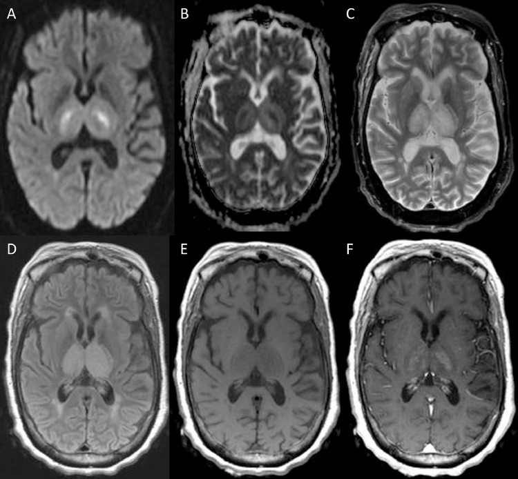
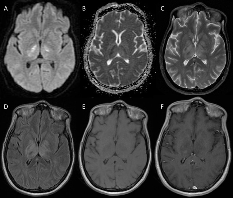
West Nile virus (WNV) is a single-stranded RNA arbovirus of Flavivirus genus that is endemic to the United States and known to cause neuroinvasive disease. Diagnosis is confirmed by the presence of WNV-specific IgM antibodies within serum or cerebrospinal fluid (CSF). Radiologically, it presents as hyperintense T2 signal within deep brain structures (ie, thalami and mid-brain) with or without cerebral peduncle and substantia nigra involvement. On diffusion-weighted imaging, restricted diffusion is reported in basal ganglia and disseminated throughout the white matter. In this report, we describe the imaging findings for 2 cases of WNV from our institution; a 56-year-old female and a 34-year-old female. Increased vigilance for WNV is warranted, particularly in immunosuppressed patients presenting with a clinical picture of viral meningoencephalitis despite initial negative magnetic resonance imaging or CSF analysis. A high suspicion for WNV disease should prompt repeat imaging or laboratory workup.
西尼罗河病毒(WNV)是黄病毒属的一种单链RNA虫媒病毒,在美国呈地方性流行,已知可引起神经侵袭性疾病。通过血清或脑脊液(CSF)中存在WNV特异性IgM抗体来确诊。在影像学上,它表现为深部脑结构(即丘脑和中脑)内T2信号增强,伴或不伴有大脑脚和黑质受累。在扩散加权成像上,基底节区有扩散受限,并在整个白质中广泛分布。在本报告中,我们描述了我们机构的2例WNV病例的影像学表现;一名56岁女性和一名34岁女性。对于WNV需要提高警惕,特别是对于尽管初始磁共振成像或脑脊液分析为阴性,但呈现病毒性脑膜脑炎临床表现的免疫抑制患者。对WNV疾病的高度怀疑应促使进行重复影像学检查或实验室检查。