Alzahrani Adel, Yadav Sumit, Gandhi Vaibhav, Lurie Alan G, Tadinada Aditya
Division of Oral and Maxillofacial Radiology, Department of Oral Health and Diagnostic Sciences, University of Connecticut School of Dental Medicine, Farmington, CT, USA.
Division of Orthodontics, University of Connecticut School of Dental Medicine, Farmington, CT, USA.
Imaging Sci Dent. 2020 Sep;50(3):245-253. doi: 10.5624/isd.2020.50.3.245. Epub 2020 Sep 16.
This study investigated the prevalence of temporomandibular joint osteoarthritis (TMJ-OA) using the Research Diagnostic Criteria for Temporomandibular Disorders image analysis criteria, assessed the severity of incidental osteoarthritic changes affecting the TMJ, and evaluated the correlations of sex and age with the prevalence and severity of TMJ-OA.
This retrospective study assessed 145 randomly selected cone-beam computed tomography scans (261 TMJs) from the authors' institutional maxillofacial radiology archive following the application of inclusion and exclusion criteria. The criteria described by Ahmad et al. were used to determine whether each TMJ was affected by OA, and the severity of the osteoarthritic changes was scored for each joint based on the method described by Alexiou et al. The chi-square, McNemar, Bhapkar chi-square, and Stuart-Maxwell chi-square tests were applied to evaluate the significance of the relationships between variables (age and sex).
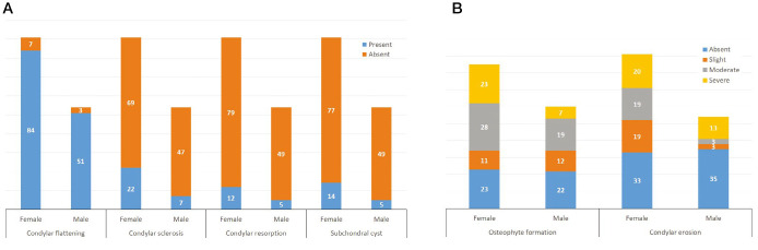
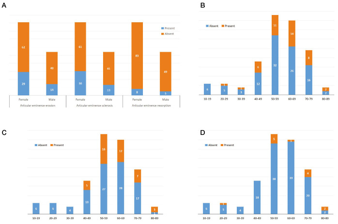
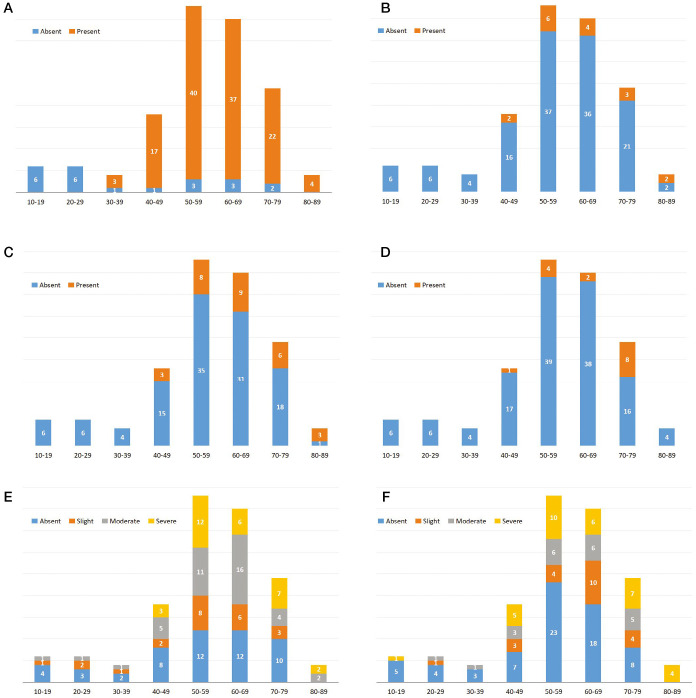
Sixteen TMJs (6.1%) had no OA, 74 (28.6%) were indeterminate for OA, and 171 (65.5%) had OA. Flattening and sclerosis were observed in 86.6% and 12.3% of cases, respectively, while resorption was observed in 7.3% of the joints. Only 21 (8.1%) of the examined TMJs had subchondral cysts. Erosion of the articular eminence was observed in 58 (22.1%) cases, while sclerosis and resorption were found in 68 (25.9%) and 16 (6.1%) TMJs, respectively.
Female patients had a higher prevalence and severity of TMJ-OA than male patients. The prevalence and severity of TMJ-OA increased with age, with peaks in the fifth and seventh decades of life.
本研究采用颞下颌关节紊乱病研究诊断标准中的影像分析标准,调查颞下颌关节骨关节炎(TMJ - OA)的患病率,评估影响颞下颌关节的偶然骨关节炎改变的严重程度,并评估性别和年龄与TMJ - OA患病率及严重程度的相关性。
本回顾性研究在应用纳入和排除标准后,从作者所在机构的颌面放射学档案中评估了145例随机选择的锥形束计算机断层扫描(261个颞下颌关节)。采用艾哈迈德等人描述的标准来确定每个颞下颌关节是否受骨关节炎影响,并根据阿列克西乌等人描述的方法对每个关节的骨关节炎改变严重程度进行评分。应用卡方检验、麦克尼马尔检验、巴普卡尔卡方检验和斯图尔特 - 麦克斯韦卡方检验来评估变量(年龄和性别)之间关系的显著性。
16个颞下颌关节(6.1%)无骨关节炎,74个(28.6%)骨关节炎情况不确定,171个(65.5%)有骨关节炎。分别在86.6%和12.3%的病例中观察到扁平及硬化,而在7.3%的关节中观察到吸收。在所检查的颞下颌关节中,只有21个(8.1%)有软骨下囊肿。在58个(22.1%)病例中观察到关节结节侵蚀,而在68个(25.9%)和16个(6.1%)颞下颌关节中分别发现硬化和吸收。
女性患者TMJ - OA的患病率和严重程度高于男性患者。TMJ - OA的患病率和严重程度随年龄增加而升高,在生命的第五和第七个十年达到峰值。