Department of Nuclear Medicine, University Hospital of Cologne, Kerpener Str. 62, 50937, Cologne, Germany.
Department of Medical Oncology, Dana-Farber Cancer Institute, Harvard Medical School, Boston, USA.
Mol Imaging Biol. 2021 Apr;23(2):277-286. doi: 10.1007/s11307-020-01546-0. Epub 2020 Oct 1.
PSMA imaging is frequently used for monitoring of androgen deprivation therapy (ADT) in prostate cancer. In a previous study, [F]-JK-PSMA-7 exhibited favorable properties for tumor localization after biochemical recurrence. In this retrospective study, we evaluated the performance of [F]-JK-PSMA-7 under ADT.
We examined the performance of [F]-JK-PSMA-7 in 70 patients (first cohort) with increasing or detectable PSA values under ADT (PSA < 2 ng/ml for 21/70 patients). We further analyzed 58 independent patients with PSA levels < 2 ng/ml under ADT, who were imaged with [Ga]PSMA-11 or [F]DCFPyL (second cohort). Finally, we compared detection rates between [F]-JK-PSMA-7, [Ga]PSMA-11, and [F]DCFPyL.
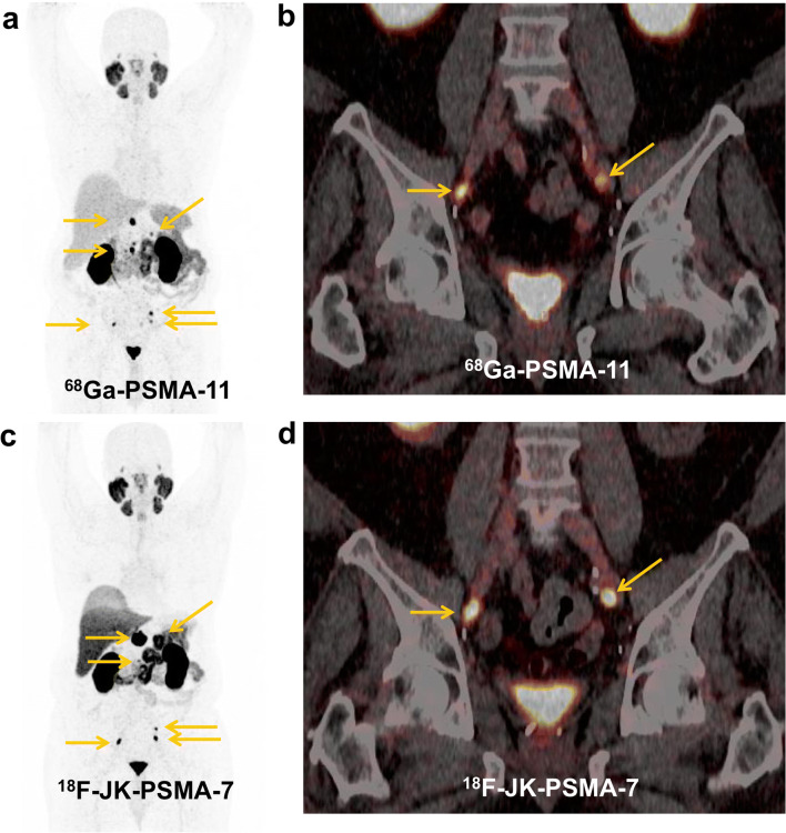
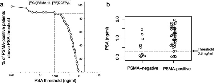
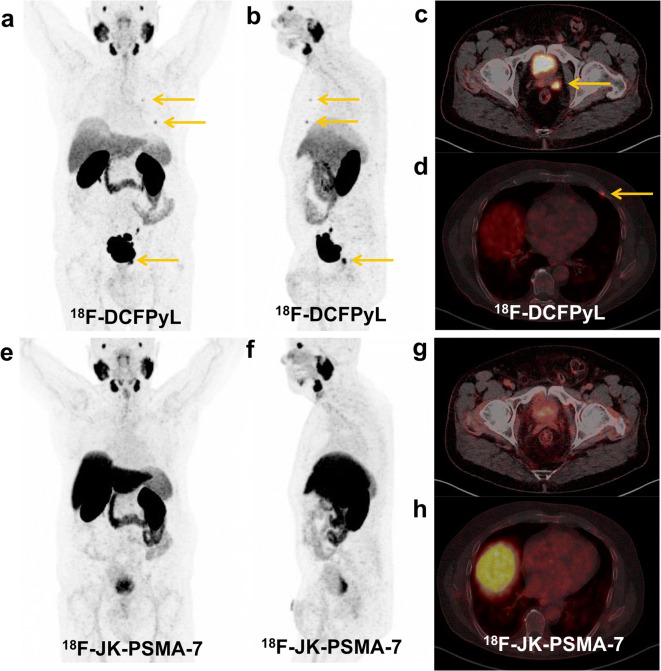
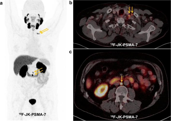
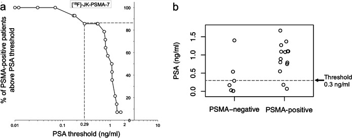
In the first cohort, we detected [F]-JK-PSMA-7-positive lesions in 63/70 patients. In patients with PSA levels ≥ 2 ng/ml, the detection rate was 100 % (49/49). In patients with PSA < 2 ng/ml, the detection rate was significantly lower (66.7 %, 14/21, p = 9.7 × 10) and dropped from 85.7 % (12/14, PSA levels between 0.3 and 2.0 ng/ml) to 28.6 % (2/7) for PSA levels < 0.3 ng/ml (p = 1.73 × 10). In the second cohort (PSA < 2 ng/ml), the detection rate was 79.3 % (46/58) for [Ga]PSMA-11 or [F]DCFPyL. Again, the detection rate was significantly higher (p = 1.1 × 10) for patients with PSA levels between 0.3 and 2.0 ng/ml (87.0 %, 40/46) relative to those with PSA levels < 0.3 ng/ml (50 %, 6/12). No significant difference was found between [F]-JK-PSMA-7 and [Ga]PSMA-11 or [F]DCFPyL in patients with PSA levels < 2 ng/ml (p = 0.4295).
[F]-JK-PSMA-7 PET showed a high detection rate in patients with PSA levels ≥ 0.3 ng/ml under ADT. The lower PSA threshold of 0.3 ng/ml for high detection rates was consistent across the three PSMA ligands. Thus, PSMA imaging is suitable for clinical follow-up of patients with increasing PSA levels under ADT.
PSMA 成像常用于监测前列腺癌的去势治疗(ADT)。在之前的一项研究中,[F]-JK-PSMA-7 在生化复发后显示出良好的肿瘤定位特性。在这项回顾性研究中,我们评估了[F]-JK-PSMA-7 在 ADT 下的性能。
我们检查了[F]-JK-PSMA-7 在 70 名 PSA 值升高或可检测的 ADT 患者(21/70 名患者 PSA<2ng/ml)中的表现。我们进一步分析了 58 名 PSA 水平<2ng/ml 的独立 ADT 患者,他们接受了[Ga]PSMA-11 或[F]DCFPyL 成像(第二队列)。最后,我们比较了[F]-JK-PSMA-7、[Ga]PSMA-11 和[F]DCFPyL 的检测率。
在第一队列中,我们在 70 名患者中的 63 名患者中检测到[F]-JK-PSMA-7 阳性病变。在 PSA 水平≥2ng/ml 的患者中,检测率为 100%(49/49)。在 PSA<2ng/ml 的患者中,检测率显著降低(66.7%,14/21,p=9.7×10),从 PSA 水平为 0.3 至 2.0ng/ml 的 85.7%(12/14)降至 PSA 水平<0.3ng/ml 的 28.6%(2/7)(p=1.73×10)。在第二队列(PSA<2ng/ml)中,[Ga]PSMA-11 或[F]DCFPyL 的检测率为 79.3%(46/58)。同样,PSA 水平在 0.3 至 2.0ng/ml 之间的患者(87.0%,40/46)的检测率明显更高(p=1.1×10),而 PSA 水平<0.3ng/ml 的患者(50%,6/12)。在 PSA<2ng/ml 的患者中,[F]-JK-PSMA-7 与[Ga]PSMA-11 或[F]DCFPyL 之间未发现显著差异(p=0.4295)。
ADT 下 PSA 水平≥0.3ng/ml 的患者中,[F]-JK-PSMA-7 PET 具有较高的检测率。三种 PSMA 配体的高检测率的 PSA 阈值为 0.3ng/ml。因此,PSMA 成像适用于 PSA 水平升高的 ADT 患者的临床随访。