Tan Li, Register Thomas C, Yammani Raghunatha R
1Section of Molecular Medicine, Department of Internal Medicine, Wake Forest School of Medicine, Winston-Salem, NC 27157, USA.
2Departments of Pathology and Comparative Medicine, Wake Forest School of Medicine, Winston-Salem, NC 27157, USA.
Aging Dis. 2020 Oct 1;11(5):1091-1102. doi: 10.14336/AD.2019.1130. eCollection 2020 Oct.
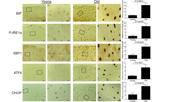
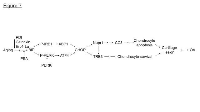
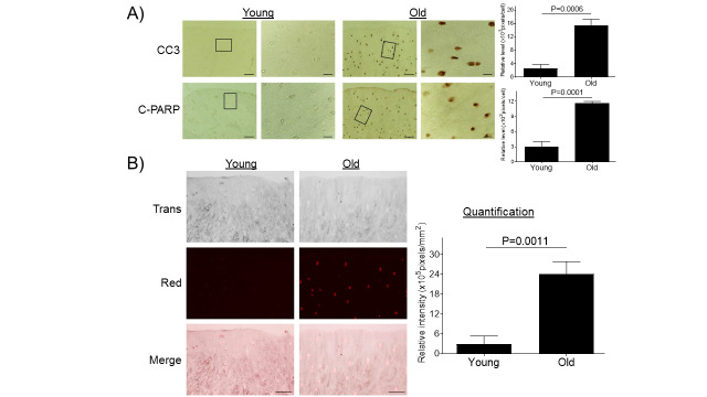
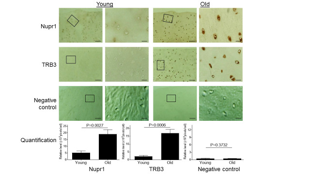
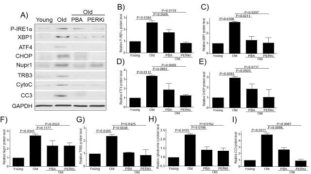
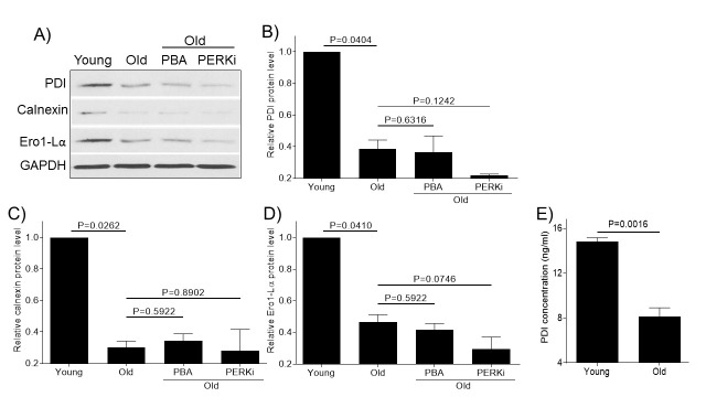
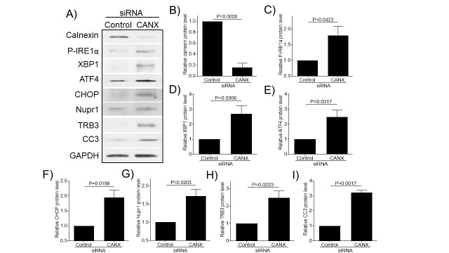
Aging is a major risk factor for the development of osteoarthritis (OA). One hallmark of aging is loss of proteostasis resulting in increased cellular stress and cell death. However, its effect on the development of OA is not clear. Here, using knee articular cartilage tissue from young and old cynomolgus monkeys (), we demonstrate that with aging there is loss of molecular chaperone expression resulting in endoplasmic reticulum (ER) stress and cell death. Chondrocytes from aged articular cartilage showed decreased expression of molecular chaperones, including protein disulfide isomerase, calnexin, and Ero1-like protein alpha, and increased immunohistochemical staining for ER stress markers (phosphorylated IRE1 alpha, spliced X-box binding protein-1, activating transcription factor 4 and C/EBP homologous protein), and apoptotic markers [cleaved caspase 3 and cleaved poly(ADP-ribose) polymerase], suggesting that decreased expression of molecular chaperone during aging induces ER stress and chondrocyte apoptosis in monkey articular cartilage. Apoptosis induced by aging-associated ER stress was further confirmed by TUNEL staining. Aged monkey cartilage also showed increased expression of nuclear protein 1 (Nupr1) and related protein-3 (TRB3), known regulators of apoptosis and cell survival pathways. Treatment of cultured monkey chondrocytes with a small molecule chemical chaperone, 4-phenylbutyric acid (PBA, a general ER stress inhibitor) or PERK Inhibitor I (an ER stress inhibitor specifically targeting the PERK branch of the unfolded protein response pathway), decreased the expression of ER stress and apoptotic markers and reduced the expression of Nupr1 and TRB3. Consistent with the above finding, knockdown of calnexin expression induces ER stress and apoptotic markers in normal human chondrocytes . Taken together, our study clearly demonstrates that aging promotes loss of proteostasis and induces ER stress and chondrocyte apoptosis in articular cartilage. Thus, restoring proteostasis using chemical/molecular chaperone or ER stress inhibitor could be a therapeutic option to treat aged-linked OA.
衰老为骨关节炎(OA)发生发展的主要风险因素。衰老的一个标志是蛋白稳态丧失,导致细胞应激增加及细胞死亡。然而,其对OA发生发展的影响尚不清楚。在此,我们利用幼年和老年食蟹猴的膝关节软骨组织,证明随着衰老,分子伴侣表达丧失,导致内质网(ER)应激和细胞死亡。老年关节软骨的软骨细胞显示分子伴侣表达降低,包括蛋白二硫键异构酶、钙连蛋白和Ero1样蛋白α,并且内质网应激标志物(磷酸化的IRE1α、剪接的X盒结合蛋白-1、激活转录因子4和C/EBP同源蛋白)以及凋亡标志物[裂解的半胱天冬酶3和裂解的聚(ADP-核糖)聚合酶]的免疫组化染色增加,提示衰老过程中分子伴侣表达降低诱导了猴关节软骨中的内质网应激和软骨细胞凋亡。衰老相关内质网应激诱导的凋亡通过TUNEL染色得到进一步证实。老年猴软骨还显示核蛋白1(Nupr1)和相关蛋白-3(TRB3)表达增加,二者为凋亡和细胞存活途径的已知调节因子。用小分子化学伴侣4-苯基丁酸(PBA,一种通用的内质网应激抑制剂)或PERK抑制剂I(一种特异性靶向未折叠蛋白反应途径中PERK分支的内质网应激抑制剂)处理培养的猴软骨细胞,可降低内质网应激和凋亡标志物的表达,并降低Nupr1和TRB3的表达。与上述发现一致,钙连蛋白表达的敲低在正常人软骨细胞中诱导内质网应激和凋亡标志物。综上所述,我们的研究清楚地表明,衰老促进关节软骨中蛋白稳态丧失,诱导内质网应激和软骨细胞凋亡。因此,使用化学/分子伴侣或内质网应激抑制剂恢复蛋白稳态可能是治疗与衰老相关OA的一种治疗选择。