Ikezu Masahiro, Edama Mutsuaki, Matsuzawa Kanta, Kaneko Fumiya, Shimizu Sohei, Hirabayashi Ryo, Kageyama Ikuo
Institute for Human Movement and Medical Sciences, Niigata University of Health and Welfare, Niigata, Japan.
Department of Anatomy, School of Life Dentistry at Niigata, Nippon Dental University, Niigata, Japan.
Orthop J Sports Med. 2020 Sep 21;8(9):2325967120952415. doi: 10.1177/2325967120952415. eCollection 2020 Sep.
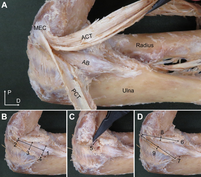
The anterior bundle (AB) of the ulnar collateral ligament is the most important structure for valgus stabilization of the elbow. However, anatomic relationships among the AB, posterior bundle (PB) of the ulnar collateral ligament, and common tendon (CT) of the flexor-pronator muscles have not been fully clarified.
To classify the AB, PB, and CT and to clarify their morphological features.
Descriptive laboratory study.
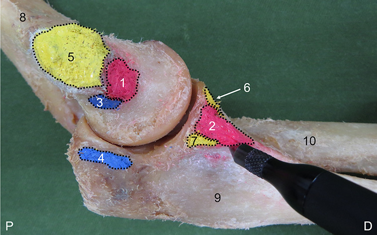
This investigation examined 56 arms from 31 embalmed Japanese cadavers. The CT investigation examined 34 arms from 23 embalmed Japanese cadavers with CTs remaining. Type classification was performed by focusing on positional relationships with surrounding structures. Morphological features measured were length, width, thickness, and footprint for the AB and PB and attachment length, thickness, and footprint for the CT.
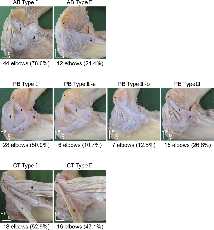
The AB was classified as type I (44 elbows; 78.6%), can be separated as a single bundle, or type II (12 elbows; 21.4%), cannot be separated from the PB and joint capsule. The PB was classified as type I (28 elbows; 50.0%), can be separated as a single bundle; type IIa (6 elbows; 10.7%), posterior edge cannot be separated; type IIb (7 elbows; 12.5%), anterior edge cannot be separated; or type III (15 elbows; 26.8%), cannot be separated from the joint capsule. The CT was classified as type I (18 elbows; 52.9%), can be separated from the AB, or type II (16 elbows; 47.1%), cannot be separated from the AB. Significant differences in frequencies of AB, PB, and CT types were identified between men and women. Morphological features were measured only for type I of each structure, and reliability was almost perfect.
These results suggest that the AB, PB, and CT each can be classified into an independent form and an unclear form. Presence of the unclear form was suggested as one factor contributing to morphological variation.
This study may provide basic information for clarifying functional roles of the AB, PB, and CT.
尺侧副韧带前束(AB)是维持肘关节外翻稳定的最重要结构。然而,尺侧副韧带前束、后束(PB)与屈肌-旋前肌总腱(CT)之间的解剖关系尚未完全阐明。
对AB、PB和CT进行分类,并阐明其形态学特征。
描述性实验室研究。
本研究检查了31具防腐处理的日本尸体的56侧上肢。CT检查了23具防腐处理的日本尸体中保留CT的34侧上肢。通过关注与周围结构的位置关系进行类型分类。测量的形态学特征包括AB和PB的长度、宽度、厚度和附着面积,以及CT的附着长度、厚度和附着面积。
AB分为I型(44例肘关节;78.6%),可分离为单束,或II型(12例肘关节;21.4%),不能与PB和关节囊分离。PB分为I型(28例肘关节;50.0%),可分离为单束;IIa型(6例肘关节;10.7%),后缘不能分离;IIb型(7例肘关节;12.5%),前缘不能分离;或III型(15例肘关节;26.8%),不能与关节囊分离。CT分为I型(18例肘关节;52.9%),可与AB分离,或II型(16例肘关节;47.1%),不能与AB分离。男性和女性在AB、PB和CT类型的频率上存在显著差异。仅对每种结构的I型测量了形态学特征,可靠性几乎完美。
这些结果表明,AB、PB和CT均可分为独立型和不明确型。不明确型的存在被认为是导致形态变异的一个因素。
本研究可为阐明AB、PB和CT的功能作用提供基础信息。