Hüneburg R, Heling D, Kaczmarek D J, van Heteren P, Olthaus M, Fimmers R, Berger M, Coch C, Lau J F, Kristiansen G, Weismüller T J, Spier I, Aretz S, Strassburg C P, Nattermann J
Department of Internal Medicine I, University Hospital Bonn, Germany.
National Center for Hereditary Tumor Syndromes, University Hospital Bonn, Germany.
Endosc Int Open. 2020 Oct;8(10):E1308-E1314. doi: 10.1055/a-1220-6699. Epub 2020 Sep 22.
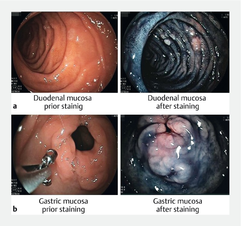
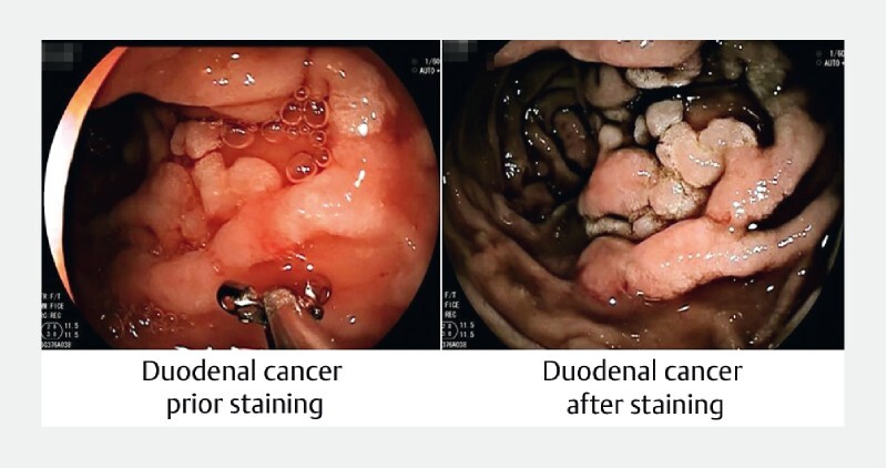
Duodenal cancer is the cancer most often seen in patients with familial adenomatous polyposis (FAP) who have undergone risk-reducing colonic surgery. Almost all patients with FAP eventually develop duodenal adenomas and risk for duodenal cancer is up to 12 % with poor prognosis. In addition, there is a rising concern regarding increased gastric cancer risk in patients with FAP. Our aim was to enhance polyp detection by using CE (CE) with the application of indigo carmine dye. We conducted a prospective, blinded study of patients with FAP undergoing endoscopic examination of the upper gastrointestinal tract. First, a standard white-light examination (WLE) was done followed by an examination performed by an endoscopist who was blinded to the previous examination, using chromoendoscopy (CE) (0.4 % indigo carmine dye). Fifty patients were included in the study. Using WLE, a median number of 13 adenomas (range 0-90) was detected compared to 23 adenomas/patient (range 0-150; < 0.0001) detected after staining, leading to a higher Spigelman stage in 16 patients (32 %; = 0.0003). CE detected significantly more larger adenomas (> 10 mm) than WLE (12 vs. 19; = 0.0391). In the gastric antral region, a median number of 0 adenomas (range 0-6) before and 0.5 adenomas (range 0-7) after staining ( = 0.0025) were detected. This prospective endoscopic trial, to our knowledge the largest in patients with FAP, showed a significant impact of CE on adenoma detection and therapeutic management in the upper gastrointestinal tract. This leads to more intensive surveillance intervals.
十二指肠癌是家族性腺瘤性息肉病(FAP)患者在接受降低风险的结肠手术后最常出现的癌症。几乎所有FAP患者最终都会发展为十二指肠腺瘤,患十二指肠癌的风险高达12%,预后较差。此外,FAP患者患胃癌风险增加也越来越受到关注。我们的目的是通过应用靛胭脂染料的胶囊内镜(CE)来提高息肉检测率。 我们对接受上消化道内镜检查的FAP患者进行了一项前瞻性、盲法研究。首先,进行标准白光检查(WLE),然后由对先前检查结果不知情的内镜医师使用色素内镜检查(CE)(0.4%靛胭脂染料)进行检查。 50名患者纳入研究。使用WLE检测到的腺瘤中位数为13个(范围0 - 90个),而染色后检测到的腺瘤为23个/患者(范围0 - 150个; <0.0001),导致16名患者(32%; =0.0003)的斯皮格尔曼分期更高。CE检测到的大于10mm的较大腺瘤明显多于WLE(12个对19个; =0.0391)。在胃窦区域,染色前腺瘤中位数为0个(范围0 - 6个),染色后为0.5个(范围0 - 7个)( =0.0025)。 据我们所知,这项前瞻性内镜试验是FAP患者中规模最大的,显示CE对上消化道腺瘤检测和治疗管理有显著影响。这导致监测间隔更加密集。