Department of Maxillofacial Surgery, University Hospital of Lübeck, Ratzeburger Allee 160, 23562, Lübeck, Germany.
Fraunhofer Research Institution for Marine Biotechnology and Cell Technology, Mönkhofer Weg 239 a, 23562, Lübeck, Germany.
Sci Rep. 2020 Oct 8;10(1):16861. doi: 10.1038/s41598-020-73941-0.
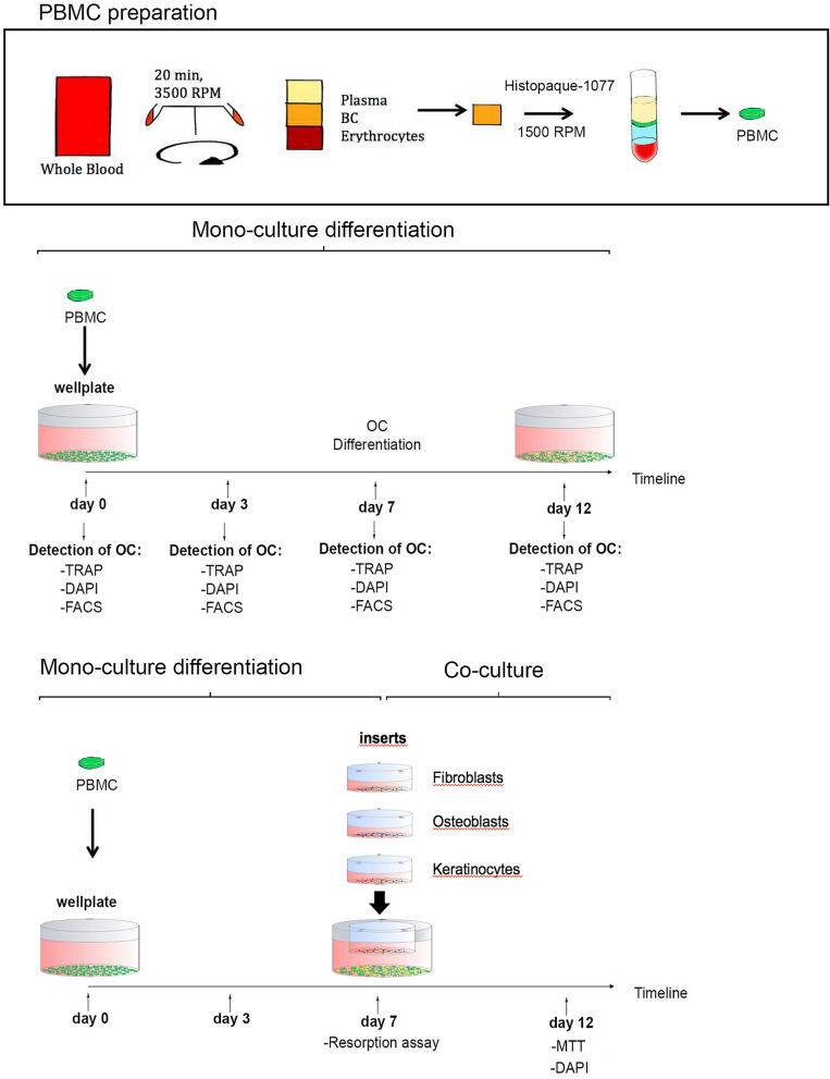
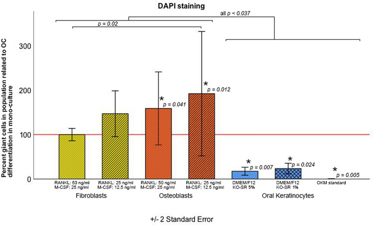
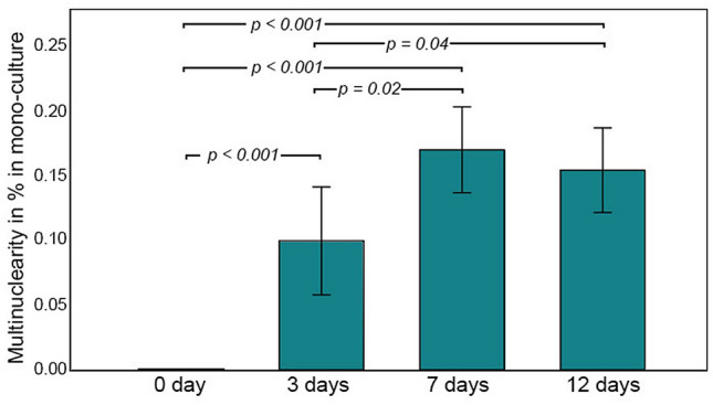
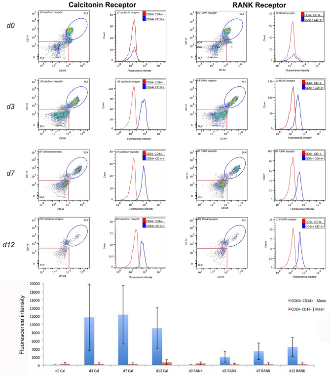
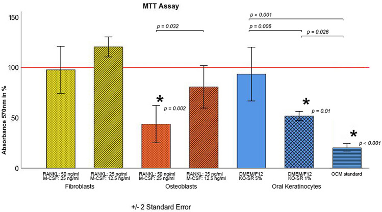
Indirect co-culture models with osteoclasts including oral cell lines may be influenced by M-CSF and RANKL in the common cell medium. Therefore, we investigated the viability and proliferation of osteoblasts (OB), fibroblasts (FB) and oral keratinocytes (OK) under stratified medium modification and assessed the differentiation of osteoclasts in each co-culture. The impact of M-CSF and RANKL in the common OC co-culture was assessed for OB, FB and OK via MTT assay via DAPI control. The multinuclearity and function of OC were evaluated by light microscopy, DAPI staining, resorption assay and FACS analysis. The PBMC showed the highest differentiation into OC after an incubation period of 7 days. Furthermore, co-culture with OB enhanced the number of differentiated multinucleated OC in comparison with monoculture, whereas co-culture with OK decreased PBMC multinuclearity and OC differentiation. FB did not influence the number of differentiated OC in a co-culture. RANKL and M-CSF reduction had no impact on OC differentiation in co-culture with FB or OB, whereas this medium modification for OK attenuated PBMC multinuclearity and OC differentiation in all approaches. Supplementation of RANKL and M-CSF can be modified for a co-culture of PBMC with FB or OB without disturbing OC differentiation. Thus, pathogenic processes of bone remodelling involving OB, OC, FB and OK in the oral cavity can be investigated thoroughly.
在含有破骨细胞的间接共培养模型中,包括口腔细胞系,可能会受到共同细胞培养基中 M-CSF 和 RANKL 的影响。因此,我们研究了在分层培养基修饰下成骨细胞 (OB)、成纤维细胞 (FB) 和口腔角质形成细胞 (OK) 的活力和增殖情况,并评估了每种共培养物中破骨细胞的分化情况。通过 MTT 分析和 DAPI 对照评估了 M-CSF 和 RANKL 在 OC 共培养中的共同作用对 OB、FB 和 OK 的影响。通过明场显微镜、DAPI 染色、吸收试验和 FACS 分析评估 OC 的多核化和功能。在孵育 7 天后,PBMC 显示出最高的 OC 分化能力。此外,与单核培养相比,与 OB 的共培养增强了分化为多核 OC 的数量,而与 OK 的共培养减少了 PBMC 的多核化和 OC 的分化。FB 在共培养中对分化 OC 的数量没有影响。RANKL 和 M-CSF 的减少对 FB 或 OB 共培养中的 OC 分化没有影响,而这种对 OK 的培养基修饰则减弱了 PBMC 的多核化和 OC 分化在所有方法中的作用。RANKL 和 M-CSF 的补充可以在不干扰 OC 分化的情况下对 PBMC 与 FB 或 OB 的共培养进行修饰。因此,可以深入研究涉及口腔中 OB、OC、FB 和 OK 的骨重塑的致病过程。