Department of Neurosurgery, School of Medicine, Collegium Medicum, University of Warmia and Mazury, Warszawska 30, 10-082, Olsztyn, Poland.
Department of Radiology, Centre of Postgraduate Medical Education, Central Clinical Hospital of Ministry of the Interior and Administration in Warsaw, Warsaw, Poland.
Sci Rep. 2020 Oct 14;10(1):17318. doi: 10.1038/s41598-020-74411-3.
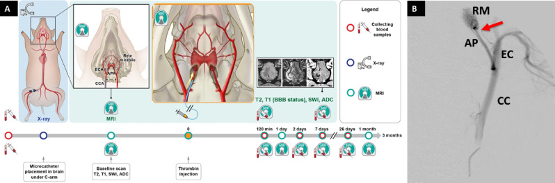
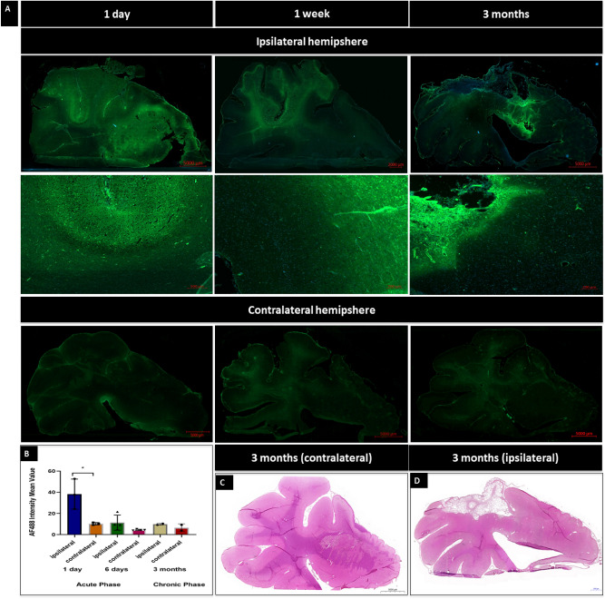
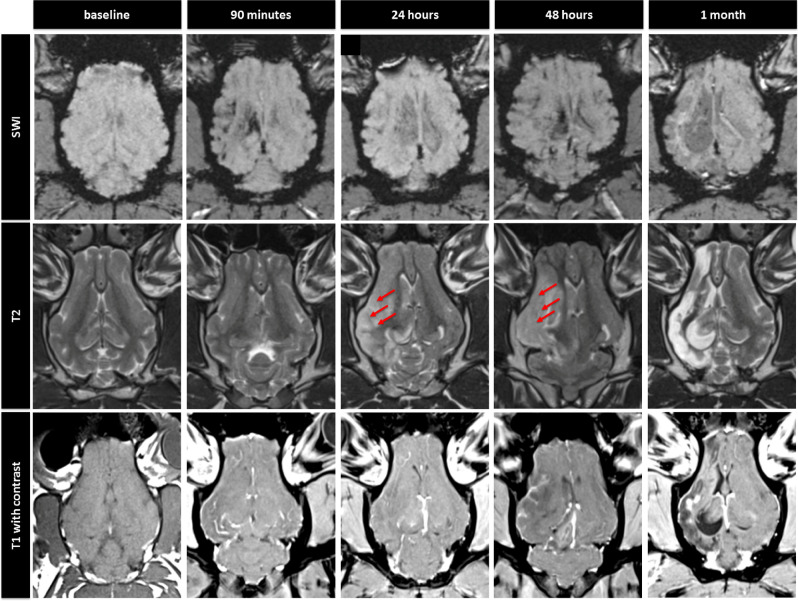
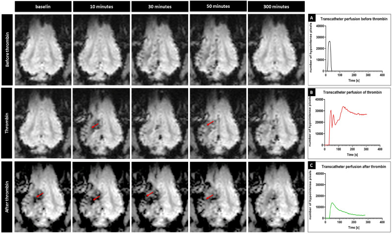
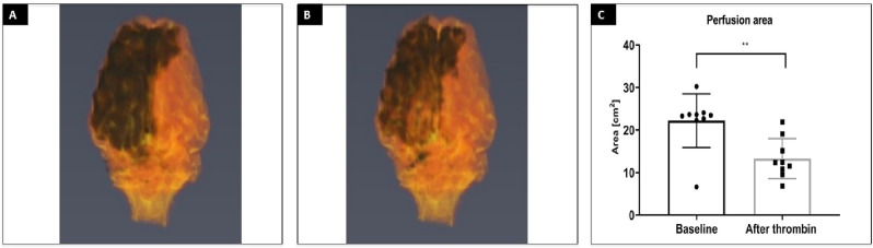
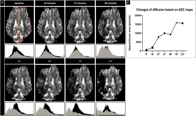
Modeling stroke in animals is essential for testing efficacy of new treatments; however, previous neuroprotective therapies, based on systemic delivery in rodents failed, exposing the need for model with improved clinical relevance. The purpose of this study was to develop endovascular approach for inducing ischemia in swine. To achieve that goal, we used intra-arterial administration of thrombin mixed with gadolinium and visualized the occlusion with real-time MRI. Placement of the microcatheter proximally to rete allowed trans-catheter perfusion of the ipsilateral hemisphere as visualized by contrast-enhanced perfusion MR scans. Dynamic T2*w MRI facilitated visualization of thrombin + Gd solution transiting through cerebral vasculature and persistent hyperintensities indicated occlusion. Area of trans-catheter perfusion dynamically quantified on representative slice before and after thrombin administration (22.20 ± 6.31 cm vs. 13.28 ± 4.71 cm respectively) indicated significantly reduced perfusion. ADC mapping showed evidence of ischemia as early as 27 min and follow-up T2w scans confirmed ischemic lesion (3.14 ± 1.41 cm). Animals developed contralateral neurological deficits but were ambulatory. Our study has overcome long lasting challenge of inducing endovascular stroke model in pig. We were able to induce stroke using minimally invasive endovascular approach and observe in real-time formation of the thrombus, blockage of cerebral perfusion and eventually stroke lesion.
在动物中进行中风模型研究对于测试新疗法的疗效至关重要;然而,以前基于啮齿动物全身给药的神经保护疗法都失败了,这表明需要一种更具临床相关性的模型。本研究旨在开发一种用于诱导猪中风的血管内方法。为了实现这一目标,我们使用动脉内给予凝血酶与钆混合,并通过实时 MRI 可视化闭塞。将微导管放置在网眼近端,可以通过对比增强灌注 MRI 扫描可视化对侧半球的经导管灌注。动态 T2*w MRI 有助于观察凝血酶+Gd 溶液通过脑血管的转运,并且持续的高信号表示闭塞。在凝血酶给药前后的代表性切片上动态定量经导管灌注的区域(分别为 22.20±6.31 cm 和 13.28±4.71 cm)表明灌注明显减少。ADC 映射显示早在 27 分钟就出现了缺血证据,并且后续的 T2w 扫描证实了缺血性病变(3.14±1.41 cm)。动物出现了对侧神经功能缺损,但仍能活动。我们的研究克服了在猪中诱导血管内中风模型的长期挑战。我们能够使用微创血管内方法诱导中风,并实时观察血栓形成、脑灌注阻塞,最终导致中风病变。