Patel Kunal S, Kejriwal Sameer, Thammachantha Samasuk, Duong Courtney, Murillo Adrian, Gordon Lynn K, Cloughesy Timothy F, Liau Linda, Yong William, Yang Isaac, Wadehra Madhuri
Department of Neurosurgery, University of California Los Angeles, Los Angeles, California, USA.
Department of Pathology and Laboratory Medicine, University of California Los Angeles, Los Angeles, California, USA.
Neurooncol Adv. 2020 Sep 8;2(1):vdaa112. doi: 10.1093/noajnl/vdaa112. eCollection 2020 Jan-Dec.
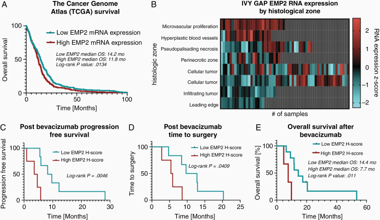
Antiangiogenic therapy with bevacizumab has failed to provide substantial gains in overall survival. Epithelial membrane protein 2 (EMP2) is a cell surface protein that has been previously shown to be expressed in glioblastoma, correlate with poor survival, and regulate neoangiogenesis in cell lines. Thus, the relationship between bevacizumab and EMP2 was investigated.
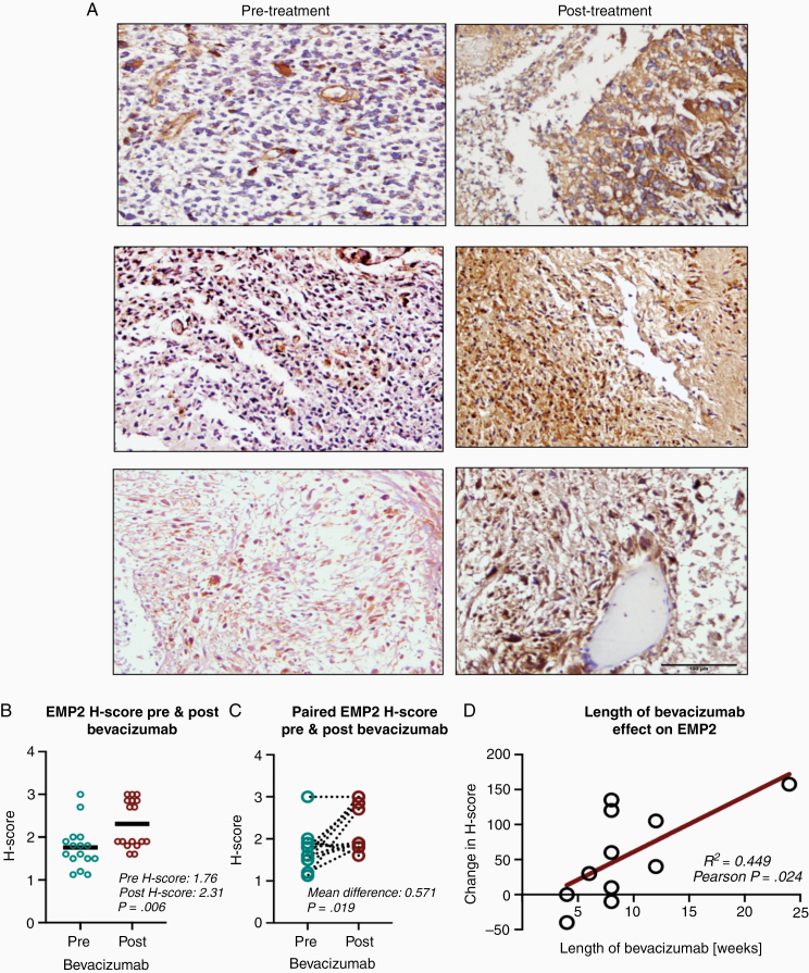
Tumor samples were obtained from 12 patients with newly diagnosed glioblastoma at 2 time points: (1) during the initial surgery and (2) during a subsequent surgery following disease recurrence post-bevacizumab treatment. Clinical characteristics and survival data from these patients were collected, and tumor samples were stained for EMP2 expression. The IVY Glioblastoma Atlas Project database was used to evaluate EMP2 expression levels in 270 samples by differing histological areas of the tumor.
Patients with high EMP2 staining at initial diagnosis had decreased progression-free and overall survival after bevacizumab (). There was increased EMP2 staining in samples obtained after bevacizumab treatment in both unpaired () and paired analyses (). This expression increase correlated with length of bevacizumab therapy ( ).
Bevacizumab treatment increased EMP2 protein expression. This increase in EMP2 correlated with reduced mean survival time post-bevacizumab therapy. We hypothesize a role of EMP2 in clinical bevacizumab resistance and as a potential antiangiogenic therapeutic target in glioblastoma.
使用贝伐单抗的抗血管生成疗法未能在总生存期方面带来显著改善。上皮膜蛋白2(EMP2)是一种细胞表面蛋白,此前已证实在胶质母细胞瘤中表达,与较差的生存率相关,并在细胞系中调节新生血管生成。因此,对贝伐单抗与EMP2之间的关系进行了研究。
从12例新诊断的胶质母细胞瘤患者在两个时间点获取肿瘤样本:(1)初次手术期间;(2)在贝伐单抗治疗后疾病复发的后续手术期间。收集这些患者的临床特征和生存数据,并对肿瘤样本进行EMP2表达染色。使用IVY胶质母细胞瘤图谱项目数据库,通过肿瘤的不同组织学区域评估270个样本中的EMP2表达水平。
初诊时EMP2染色高的患者在接受贝伐单抗治疗后的无进展生存期和总生存期缩短()。在贝伐单抗治疗后获得的样本中,未配对分析()和配对分析()中EMP2染色均增加。这种表达增加与贝伐单抗治疗的持续时间相关()。
贝伐单抗治疗增加了EMP2蛋白表达。EMP2的这种增加与贝伐单抗治疗后平均生存时间缩短相关。我们推测EMP2在临床贝伐单抗耐药中起作用,并作为胶质母细胞瘤潜在的抗血管生成治疗靶点。