Department of Nuclear Medicine, Military Institute of Medicine, 128 Szaserów St, 04-141, Warsaw, Poland.
Affidea Mazovian PET/CT Medical Centre, 128 Szaserów St, 04-349, Warsaw, Poland.
Int J Cardiovasc Imaging. 2021 Mar;37(3):1097-1104. doi: 10.1007/s10554-020-02056-4. Epub 2020 Oct 15.
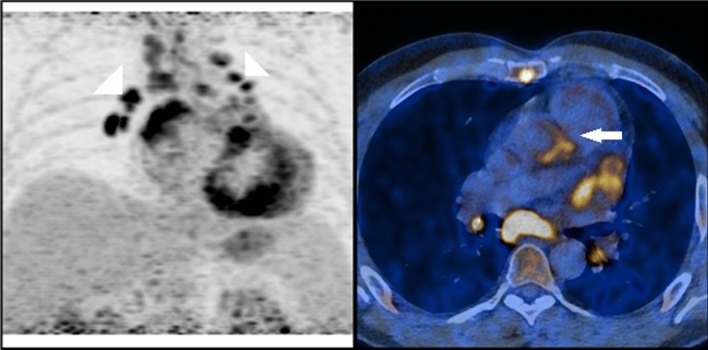
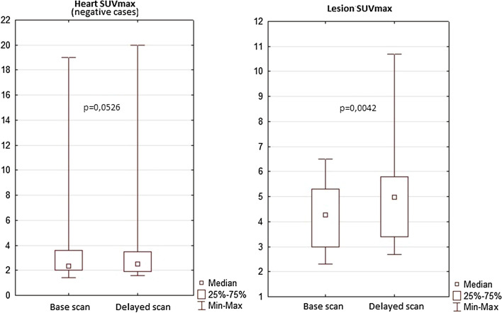
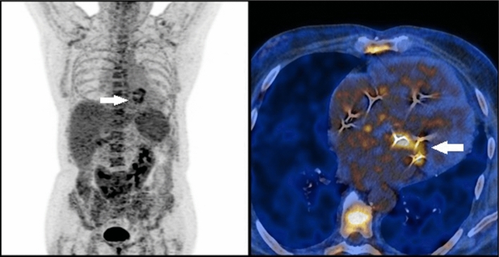
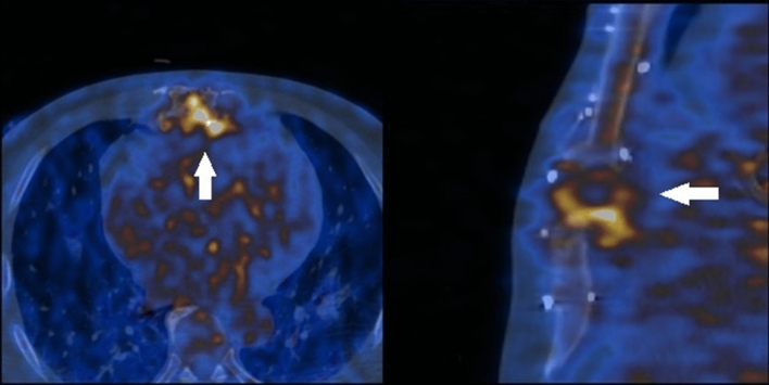
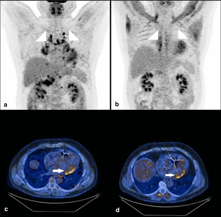
The aim of the study was to assess the feasibility of FDG PET in cardiac inflammation with a particular focus on the delayed scan. Thirty-five consecutive FDG PET scans of patients with suspected or confirmed cardiac inflammation were retrospectively reviewed. The patients were referred for PET because of endocarditis (n = 16) or sarcoidosis (n = 19). Among them four patients had two consecutive for follow up and treatment control (two patients with sarcoidosis, two with endocarditis). In all of the cases a standard head to mid-thigh scan was performed 45-60 min after FDG injection as well as a delayed heart scan 1 h after the standard imaging was performed. FDG PET confirmed active inflammation in 10 out of 35 scans. Delayed scans in positive cases showed SUVmax value increase, but did not have an impact on the result, neither they did in negative cases-no significant differences between standard and delayed scan were found. Interestingly in 5 out of 14 cases with suspected endocarditis PET revealed the extracardiac inflammation focus, thus changing initial diagnosis. FDG PET also indicated which prosthesis caused inflammation if there were many. In the sarcoidosis group the aim was to confirm or exclude heart involvement (13 scans) or to assess the response to the steroid therapy (6 scans) in patients with previously confirmed sarcoidosis. PET revealed active heart disease in 3 initial scans, and 1 follow up scan. FDG PET is a valuable imaging method for the cardiac inflammation assessment. It adequately localises the active inflammation site. Also, since it is a whole-body scan it may detect the extracardiac inflammation foci, which in some cases may change the initial diagnosis. In our study the delayed scans showed no added value.
这项研究的目的是评估 FDG PET 在心脏炎症中的可行性,特别关注延迟扫描。回顾性分析了 35 例疑似或确诊为心脏炎症的连续 FDG PET 扫描患者。这些患者因感染性心内膜炎(n=16)或结节病(n=19)而接受 PET 检查。其中 4 例患者因随访和治疗控制需要进行了两次连续扫描(2 例结节病,2 例感染性心内膜炎)。所有患者在 FDG 注射后 45-60 分钟进行标准从头到大腿扫描,并在标准成像后 1 小时进行延迟心脏扫描。FDG PET 在 35 例扫描中证实了 10 例活跃炎症。阳性病例的延迟扫描显示 SUVmax 值增加,但对结果没有影响,阴性病例也没有影响——标准扫描和延迟扫描之间没有发现显著差异。有趣的是,在 14 例疑似感染性心内膜炎的病例中,有 5 例 PET 显示了心脏外炎症灶,从而改变了最初的诊断。FDG PET 还表明,如果有多个假体,哪个假体引起了炎症。在结节病组中,目的是确认或排除心脏受累(13 例扫描),或评估先前确诊的结节病患者对类固醇治疗的反应(6 例扫描)。PET 在 3 例初始扫描和 1 例随访扫描中发现了活动性心脏病。FDG PET 是评估心脏炎症的一种有价值的成像方法。它能准确地定位活跃的炎症部位。此外,由于它是全身扫描,它可能会检测到心脏外炎症灶,在某些情况下可能会改变最初的诊断。在我们的研究中,延迟扫描没有显示出额外的价值。