• 文献检索
  • 文档翻译
  • 深度研究
  • 学术资讯
  • Suppr Zotero 插件Zotero 插件
  • 邀请有礼
  • 套餐&价格
  • 历史记录
应用&插件
Suppr Zotero 插件Zotero 插件浏览器插件Mac 客户端Windows 客户端微信小程序
定价
高级版会员购买积分包购买API积分包
服务
文献检索文档翻译深度研究API 文档MCP 服务
关于我们
关于 Suppr公司介绍联系我们用户协议隐私条款
关注我们

Suppr 超能文献

核心技术专利:CN118964589B侵权必究
粤ICP备2023148730 号-1Suppr @ 2026

文献检索

告别复杂PubMed语法,用中文像聊天一样搜索,搜遍4000万医学文献。AI智能推荐,让科研检索更轻松。

立即免费搜索

文件翻译

保留排版,准确专业,支持PDF/Word/PPT等文件格式,支持 12+语言互译。

免费翻译文档

深度研究

AI帮你快速写综述,25分钟生成高质量综述,智能提取关键信息,辅助科研写作。

立即免费体验

模拟轴突起始段中的蛋白质-蛋白质相互作用,以了解它们对动作电位起始的潜在影响。

Modeling protein-protein interactions in axon initial segment to understand their potential impact on action potential initiation.

作者信息

Bhardwaj Piyush, Kulasiri Don, Samarasinghe Sandhya

机构信息

Centre of Advanced Computational Solutions (C-fACS); Department of Molecular Biosciences, Lincoln University, Christchurch, New Zealand.

Centre of Advanced Computational Solutions (C-fACS), Lincoln University, Christchurch, New Zealand.

出版信息

Neural Regen Res. 2021 Apr;16(4):700-706. doi: 10.4103/1673-5374.295332.

DOI:10.4103/1673-5374.295332
PMID:33063731
原文链接:https://pmc.ncbi.nlm.nih.gov/articles/PMC8067952/
Abstract

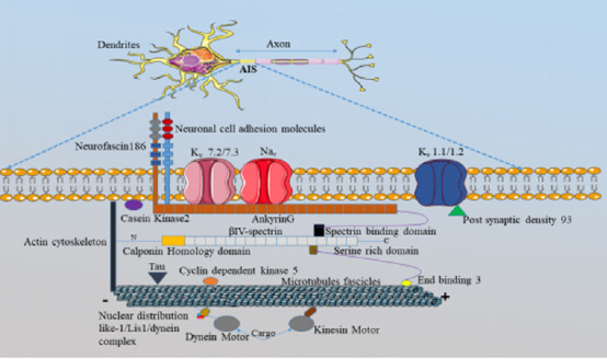
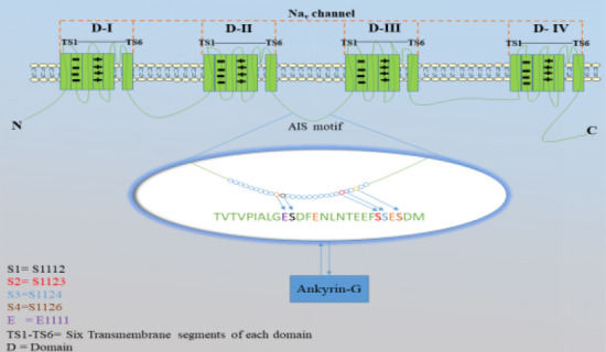
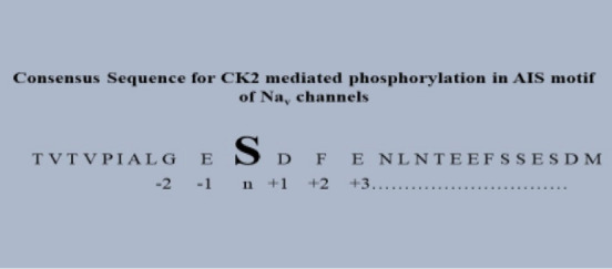
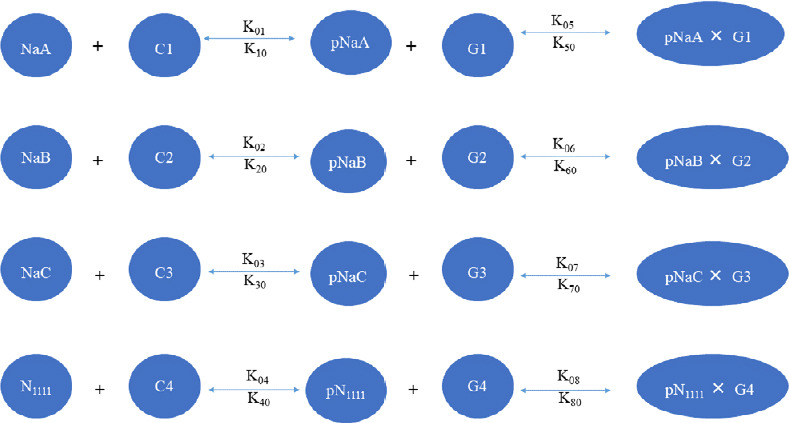
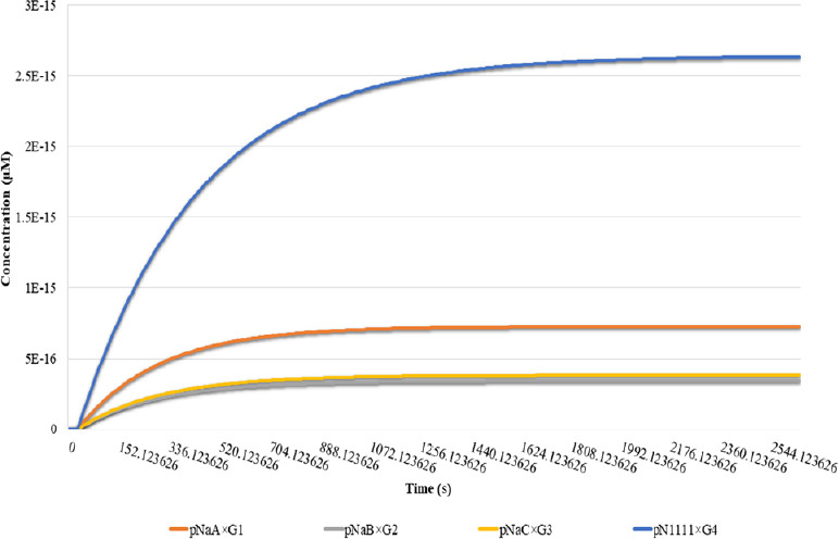
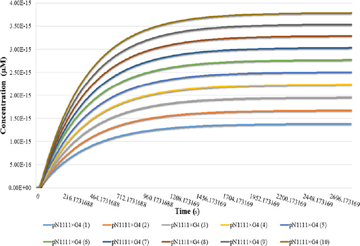
The axon initial segment (AIS) region is crucial for action potential initiation due to the presence of high-density AIS protein voltage-gated sodium channels (Na). Na channels comprise several serine residues responsible for the recruitment of Na channels into the structure of AIS through interactions with ankyrin-G (AnkG). In this study, a series of computational experiments are performed to understand the role of AIS proteins casein kinase 2 and AnkG on Na channel recruitment into the AIS. The computational simulation results using Virtual cell software indicate that Na channels with all serine sites available for phosphorylation bind to AnkG with strong affinity. At the low initial concentration of AnkG and casein kinase 2, the concentration of Na channels reduces significantly, suggesting the importance of casein kinase 2 and AnkG in the recruitment of Na channels.

摘要

轴突起始段(AIS)区域对于动作电位的起始至关重要,这是因为存在高密度的AIS蛋白电压门控钠通道(Na)。钠通道包含几个丝氨酸残基,这些残基通过与锚蛋白G(AnkG)相互作用负责将钠通道招募到AIS结构中。在本研究中,进行了一系列计算实验以了解AIS蛋白酪蛋白激酶2和AnkG在钠通道招募到AIS中的作用。使用虚拟细胞软件的计算模拟结果表明,所有丝氨酸位点都可用于磷酸化的钠通道与AnkG具有很强的亲和力。在AnkG和酪蛋白激酶2的初始浓度较低时,钠通道的浓度显著降低,这表明酪蛋白激酶2和AnkG在钠通道招募中的重要性。

https://cdn.ncbi.nlm.nih.gov/pmc/blobs/22eb/8067952/b1c7cf106aa4/NRR-16-700-g016.jpg
https://cdn.ncbi.nlm.nih.gov/pmc/blobs/22eb/8067952/f69c284e72b5/NRR-16-700-g001.jpg
https://cdn.ncbi.nlm.nih.gov/pmc/blobs/22eb/8067952/fbd860237be5/NRR-16-700-g002.jpg
https://cdn.ncbi.nlm.nih.gov/pmc/blobs/22eb/8067952/88071b7f618c/NRR-16-700-g003.jpg
https://cdn.ncbi.nlm.nih.gov/pmc/blobs/22eb/8067952/4111907fe0a8/NRR-16-700-g004.jpg
https://cdn.ncbi.nlm.nih.gov/pmc/blobs/22eb/8067952/2ecd9b302f33/NRR-16-700-g005.jpg
https://cdn.ncbi.nlm.nih.gov/pmc/blobs/22eb/8067952/717b5d371481/NRR-16-700-g014.jpg
https://cdn.ncbi.nlm.nih.gov/pmc/blobs/22eb/8067952/94f5e285b781/NRR-16-700-g015.jpg
https://cdn.ncbi.nlm.nih.gov/pmc/blobs/22eb/8067952/b1c7cf106aa4/NRR-16-700-g016.jpg

相似文献

1
Modeling protein-protein interactions in axon initial segment to understand their potential impact on action potential initiation.模拟轴突起始段中的蛋白质-蛋白质相互作用,以了解它们对动作电位起始的潜在影响。
Neural Regen Res. 2021 Apr;16(4):700-706. doi: 10.4103/1673-5374.295332.
2
An Ankyrin-G N-terminal Gate and Protein Kinase CK2 Dually Regulate Binding of Voltage-gated Sodium and KCNQ2/3 Potassium Channels.锚蛋白G的N端门控和蛋白激酶CK2双重调节电压门控钠通道及KCNQ2/3钾通道的结合
J Biol Chem. 2015 Jul 3;290(27):16619-32. doi: 10.1074/jbc.M115.638932. Epub 2015 May 21.
3
Ankyrin G Membrane Partners Drive the Establishment and Maintenance of the Axon Initial Segment.锚蛋白G膜伴侣驱动轴突起始段的建立和维持。
Front Cell Neurosci. 2017 Jan 26;11:6. doi: 10.3389/fncel.2017.00006. eCollection 2017.
4
Tetrodotoxin-resistant voltage-gated sodium channel Nav 1.8 constitutively interacts with ankyrin G.抗河豚毒素电压门控钠通道Nav 1.8与锚蛋白G持续相互作用。
J Neurochem. 2014 Oct;131(1):33-41. doi: 10.1111/jnc.12785. Epub 2014 Jun 27.
5
Ankyrin-G directly binds to kinesin-1 to transport voltage-gated Na+ channels into axons.锚蛋白 G 直接结合驱动蛋白-1 将电压门控钠离子通道运输到轴突中。
Dev Cell. 2014 Jan 27;28(2):117-31. doi: 10.1016/j.devcel.2013.11.023. Epub 2014 Jan 9.
6
CK2-regulated schwannomin-interacting protein IQCJ-SCHIP-1 association with AnkG contributes to the maintenance of the axon initial segment.细胞周期蛋白依赖性激酶2(CK2)调控的与施万omin相互作用的蛋白IQCJ - SCHIP - 1与锚蛋白G(AnkG)的结合有助于维持轴突起始段。
J Neurochem. 2015 Aug;134(3):527-37. doi: 10.1111/jnc.13158. Epub 2015 Jun 22.
7
Crystal structure of Ankyrin-G in complex with a fragment of Neurofascin reveals binding mechanisms required for integrity of the axon initial segment.锚蛋白 G 与神经束蛋白片段复合物的晶体结构揭示了维持轴突起始段完整性所需的结合机制。
J Biol Chem. 2022 Sep;298(9):102272. doi: 10.1016/j.jbc.2022.102272. Epub 2022 Jul 16.
8
Loss of Neurofascin-186 Disrupts Alignment of AnkyrinG Relative to Its Binding Partners in the Axon Initial Segment.神经束蛋白-186的缺失会破坏锚蛋白G相对于其在轴突起始段结合伙伴的排列。
Front Cell Neurosci. 2019 Jan 22;13:1. doi: 10.3389/fncel.2019.00001. eCollection 2019.
9
Ca entry through Na channels generates submillisecond axonal Ca signaling.钙通过钠通道进入产生亚毫秒级轴突钙信号。
Elife. 2020 Jun 17;9:e54566. doi: 10.7554/eLife.54566.
10
Ankyrin G restricts ion channel diffusion at the axonal initial segment before the establishment of the diffusion barrier.锚蛋白 G 在轴突起始段扩散屏障建立之前限制离子通道在该部位的扩散。
J Cell Biol. 2010 Oct 18;191(2):383-95. doi: 10.1083/jcb.201003042.

本文引用的文献

1
A Brief History of Simulation Neuroscience.模拟神经科学简史
Front Neuroinform. 2019 May 7;13:32. doi: 10.3389/fninf.2019.00032. eCollection 2019.
2
Reconsideration of Amyloid Hypothesis and Tau Hypothesis in Alzheimer's Disease.重新审视阿尔茨海默病中的淀粉样蛋白假说和tau蛋白假说。
Front Neurosci. 2018 Jan 30;12:25. doi: 10.3389/fnins.2018.00025. eCollection 2018.
3
Axonal Membranes and Their Domains: Assembly and Function of the Axon Initial Segment and Node of Ranvier.轴突膜及其结构域:轴突起始段和郎飞结的组装与功能
Front Cell Neurosci. 2017 May 9;11:136. doi: 10.3389/fncel.2017.00136. eCollection 2017.
4
PPARgamma agonists rescue increased phosphorylation of FGF14 at S226 in the Tg2576 mouse model of Alzheimer's disease.在阿尔茨海默病的Tg2576小鼠模型中,过氧化物酶体增殖物激活受体γ(PPARγ)激动剂可挽救FGF14在S226位点磷酸化增加的现象。
Exp Neurol. 2017 Sep;295:1-17. doi: 10.1016/j.expneurol.2017.05.005. Epub 2017 May 15.
5
Structural and Functional Plasticity at the Axon Initial Segment.轴突起始段的结构与功能可塑性
Front Cell Neurosci. 2016 Oct 25;10:250. doi: 10.3389/fncel.2016.00250. eCollection 2016.
6
Axon Initial Segment Cytoskeleton: Architecture, Development, and Role in Neuron Polarity.轴突起始段细胞骨架:结构、发育及其在神经元极性中的作用
Neural Plast. 2016;2016:6808293. doi: 10.1155/2016/6808293. Epub 2016 Jul 17.
7
Acetylated tau destabilizes the cytoskeleton in the axon initial segment and is mislocalized to the somatodendritic compartment.乙酰化tau蛋白会破坏轴突起始段的细胞骨架,并错误定位于树突体部分。
Mol Neurodegener. 2016 Jun 29;11(1):47. doi: 10.1186/s13024-016-0109-0.
8
Amyloid-β plaques disrupt axon initial segments.淀粉样β蛋白斑块会破坏轴突起始段。
Exp Neurol. 2016 Jul;281:93-8. doi: 10.1016/j.expneurol.2016.04.018. Epub 2016 Apr 21.
9
Neuron Morphology Influences Axon Initial Segment Plasticity.神经元形态影响轴突起始段可塑性。
eNeuro. 2016 Feb 13;3(1). doi: 10.1523/ENEURO.0085-15.2016. eCollection 2016 Jan-Feb.
10
Nanoscale Architecture of the Axon Initial Segment Reveals an Organized and Robust Scaffold.轴突起始段的纳米级结构揭示了一种有序而坚固的支架。
Cell Rep. 2015 Dec 29;13(12):2781-93. doi: 10.1016/j.celrep.2015.11.051. Epub 2015 Dec 17.